The fundus of the eye refers to the inner surface at the back of the eyeball that can be seen during a clinical examination. It is one of the most crucial areas in ophthalmology as it allows doctors to directly observe blood vessels, the retina, the optic nerve, and the macula structures that reveal much about both the eye and overall health.
Conditions like glaucoma, diabetic retinopathy, and macular degeneration are often first detected through a fundus exam. Understanding the fundus of the eye, its anatomy, and how it is evaluated through tests such as ophthalmoscopy or fundus photography helps patients appreciate the importance of regular screening for eye health.
Introduction: Why Understanding the Fundus Matters
The fundus of the eye is the only part of the human body where blood vessels and nerve tissues can be directly viewed without invasive procedures. This makes it a key diagnostic window for detecting eye diseases and systemic conditions such as diabetes or hypertension.
The eye fundus definition includes structures like the retina, macula, fovea, optic disc, and the retinal blood vessels, all of which contribute to clear vision. The fundus exam overview involves inspecting these parts to ensure their health and identify abnormalities. Modern fundus photography introduction techniques now allow digital imaging of the retina, helping track disease progression accurately over time.
Understanding the Fundus of the Eye
Definition and Anatomy
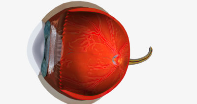
The fundus of the eye refers to the interior back surface of the eye, visible through the pupil using an ophthalmoscope. It includes key structures such as:
- Retina: The light-sensitive layer that captures images.
- Macula and Fovea: The central area responsible for detailed and colour vision.
- Optic Disc and Cup: The point where the optic nerve exits the eye, transmitting signals to the brain.
- Choroid Blood Vessels: The vascular layer supplying oxygen and nutrients to the retina.
The eye fundus anatomy is best observed using a fundus camera or ophthalmoscope, providing a detailed look at the retinal fundus and associated structures.
Function of the Fundus
The function of the fundus of the eye is essential for sight. Light enters through the cornea and lens, focusing onto the retina located in the fundus. Here, retinal photoreceptors convert light into electrical impulses transmitted through optic nerve signals to the brain for visual processing.
A healthy fundus ensures clear, sharp vision, while any structural or vascular abnormality can lead to impaired sight or blindness. Thus, assessing the fundus provides vital insights into both local eye health and systemic well-being.
Normal Appearance of the Fundus
Optic Disc and Cup
In a normal fundus of the eye, the optic disc appears round or slightly oval, with a yellow-pink hue and well-defined edges. The optic cup-to-disc ratio typically occupies less than one-third of the disc’s diameter.
The ISNT rule Inferior, Superior, Nasal, Temporal helps clinicians assess rim thickness, which is thinner temporally and thicker inferiorly. Any deviation from this pattern could indicate glaucoma or optic nerve damage.
Retinal Blood Vessels & Quadrants
Normal retinal vessels include arteries and veins branching out from the optic disc. Arteries are narrower and lighter, while veins appear broader and darker.
These vessels divide into four retinal quadrants, supplying the upper and lower hemispheres. The macula location lies about two disc diameters away from the disc, appearing as a dusky, avascular area vital for central vision.
Macula and Fovea
The macula is the region of highest visual resolution, and its centre, the fovea, is responsible for sharp central vision. The normal macula appearance is slightly darker due to densely packed photoreceptors. When assessing the fundus of the eye for normal appearance, a healthy macula and clear foveal reflex are key indicators of good retinal function.
How the Fundus of the Eye Is Examined
Fundoscopic Examination (Ophthalmoscopy)
The fundus exam procedure, or ophthalmoscopy, allows doctors to view internal eye structures directly. In direct ophthalmoscopy, a handheld ophthalmoscope gives a magnified, narrow view of the retina.
Indirect ophthalmoscopy, performed with a head-mounted lens system, provides a wider and stereoscopic view. The dilated eye exam ensures full visibility of the retina, helping detect conditions like glaucoma and macular degeneration early.
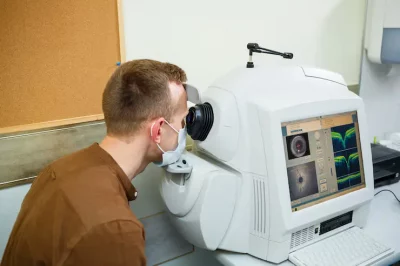
Fundus Photography and Digital Imaging
Fundus photography test captures detailed images of the retina using a wide-field fundus camera. This digital retinal imaging method enables permanent documentation of retinal health, aiding long-term monitoring.
During the retina photo test, the pupils may be dilated, and patients are asked to focus on a light while images are captured. This non-invasive eye imaging method is accurate, quick, and painless.
Other Imaging Techniques
Beyond photography, optical coherence tomography (OCT) and ultra-wide-field imaging provide high-resolution cross-sectional images of the retina, helping diagnose subtle or early-stage abnormalities in the eye fundus.
Conditions Detected Through Fundus Examination
A fundus examination of the eye can detect a wide range of diseases, including:
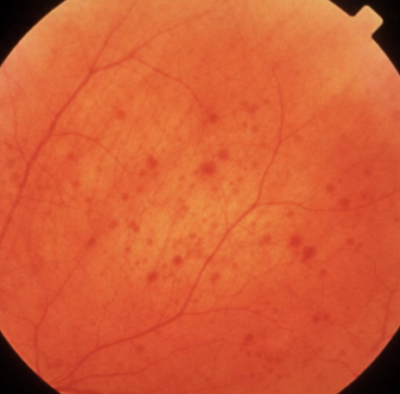
- Diabetic Retinopathy: Characterised by microaneurysms, haemorrhages, and swelling in retinal blood vessels.
- Macular Degeneration (AMD): Presence of drusen or pigment changes in the macula.
- Glaucoma: Noted by increased optic nerve cupping and rim thinning.
- Retinal Vascular Diseases: Such as arterial or venous occlusions.
- Retinal Detachment: Identified by tears, fluid, or lifted retinal tissue.
- Optic Nerve Conditions: Including optic atrophy and papilledema.
- Systemic Conditions: Diabetes, hypertension, and endocarditis can manifest visibly in the fundus of the eye.
Early detection through diabetic retinopathy screening and macular degeneration detection significantly reduces the risk of vision loss.
When to Get a Fundus Exam
Regular fundus examination of the eye is vital for everyone, especially individuals with chronic diseases or visual symptoms. High-risk groups include those with diabetes, hypertension, severe myopia, uveitis, or a family history of retinal disorders.
The fundus exam frequency varies, but is typically annual for diabetic or hypertensive patients. Understanding when a fundus exam is needed can help in early intervention and long-term visual preservation.
Preparing for Your Fundus Exam & What to Expect
During fundus exam preparation, patients are usually given dilating drops to widen the pupils for better visibility. Temporary blurred vision and light sensitivity may occur. You can bring sunglasses and arrange transport if necessary.
Knowing how to prepare for the eye fundus test ensures comfort. The process is brief and painless, and understanding what to expect during a fundus exam helps ease anxiety before the appointment.
Risks and Side Effects of Fundus Eyes
The fundus exam side effects are minimal. Temporary light sensitivity or mild blurred vision are common after pupil dilation.
Rarely, individuals may experience allergic reactions to drops or mild discomfort. In general, the risks of fundus photography are extremely low, and the test is safe, painless, and essential for preserving long-term eye health.
Conclusion: Protect Your Vision Through Regular Fundus Exams
The fundus of the eye provides a direct view of critical structures that reflect both ocular and systemic health. A normal fundus of the eye shows healthy vessels, a clear macula, and a well-defined optic disc.
Regular fundus exams using modern imaging technologies allow early diagnosis of serious conditions, helping you protect your vision effectively. Prioritising these examinations supports early detection, intervention, and lifelong eye health.