The cornea is often called the eye’s “window” because it allows light to enter and plays a central role in vision. This transparent, dome-shaped surface covers the front of the eye and bends light to help it focus on the retina.
Understanding the anatomy of the cornea is essential to appreciating how delicate and vital it is. Each of the layers of the cornea contributes to maintaining clarity, shape, and overall eye health. Damage or disease affecting any layer of the eye can impair vision, making corneal care and protection crucial for long-term ocular well-being.
What is the Cornea?
The cornea is the transparent outermost layer of the eye that acts as a barrier and a lens. It covers the iris, pupil, and anterior chamber. Structurally, it is part of the layers of the eye, which include the cornea, sclera, choroid, retina, and other internal structures. The layers of the cornea are specially designed to maintain transparency and refractive accuracy.
Together, these layers help focus light precisely onto the retina. Unlike other tissues, the cornea is avascular, meaning it contains no blood vessels, which allows light to pass through unobstructed. Instead, it receives nourishment from tears and the aqueous humour.
The Function of the Cornea in Eye Health
The cornea performs two essential functions: refraction and protection. It provides nearly two-thirds of the eye’s total focusing power and acts as a shield against dust, germs, and harmful UV rays. The 6 layers of the cornea work together to maintain a clear optical surface.
This transparency ensures that light enters the eye smoothly and reaches the retina without scattering. In addition, the cornea helps protect the inner layers of the eye from mechanical injury and infection. Each corneal layer, from the epithelium to the endothelium, plays a unique role in preserving both structure and function.
The Anatomy of the Cornea
Layers of the Cornea
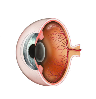
The cornea is made up of multiple layers that work in harmony to provide optical clarity and protection. Traditionally, anatomists identified 5 layers of the cornea, but modern research has recognised a sixth layer, called Dua’s layer, leading to the term 6 layers of cornea.
A diagram of the corneal layers or a histology illustration of the corneal layers can help visualise this structure. Each layer has a distinct composition and function, contributing to the cornea’s strength, resilience, and transparency.
1. Epithelium
The epithelium is the outermost layer of the cornea and serves as the eye’s first line of defence against environmental threats such as dust, debris, and microorganisms. It also absorbs oxygen and nutrients from tears, distributing them to the deeper layers of the eye.
Histologically, this layer consists of several tightly packed cell layers that regenerate rapidly to heal minor abrasions. The epithelium is essential for maintaining smooth optical quality and protecting the inner layers.
2. Bowman’s Layer
Located beneath the epithelium, Bowman’s layer is a thin but tough sheet of collagen fibres. It provides the cornea with additional strength and stability, helping it maintain shape and resist injury.
Although this layer cannot regenerate after damage, its structural integrity is vital in preventing deeper injury to the stroma. This layer plays an important role in the biomechanical strength of the corneal layers.
3. Stroma
The stroma forms about 90% of the cornea’s total thickness, making it the largest of the layers of the eye within the cornea. It is composed of regularly arranged collagen fibres and keratocytes, which maintain transparency and strength.
Any disruption to this arrangement, such as in scarring or infection, can cause light scattering and reduce visual clarity. The stroma’s water content must remain balanced, regulated by the deeper endothelial layer.
4. Descemet’s Membrane
Descemet’s membrane is a thin but highly elastic layer located beneath the stroma. It acts as a protective barrier and supports the endothelium.
This layer regenerates efficiently after minor injuries and helps maintain corneal flexibility and stability. Despite being one of the thinnest layers of the cornea, it is essential in maintaining corneal resilience and protection against infections or inflammation.
5. Endothelium
The endothelium is the innermost layer of the cornea and is responsible for maintaining fluid balance. Its cells act like a pump, preventing excess water from entering the stroma and maintaining corneal transparency.
Damage to this layer can cause corneal swelling (oedema), leading to blurred vision. The layers of cornea histology show that endothelial cells do not regenerate easily, making their preservation crucial for long-term eye health.
Common Corneal Conditions
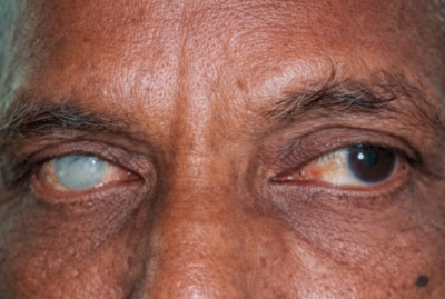
Diseases affecting the corneal layers can lead to vision problems ranging from mild discomfort to severe vision loss. Common conditions include keratitis, corneal ulcers, and corneal dystrophies. Infections or injuries may damage the epithelial layer, while deeper issues such as keratoconus affect the stroma’s structure, leading to thinning and distortion.
Conditions such as Fuchs’ endothelial dystrophy impact the endothelium, leading to fluid buildup and blurred vision. Using a layer of the eye diagram helps doctors identify which part of the cornea is affected and tailor treatment accordingly.
How to Keep Your Cornea Healthy
Maintaining corneal health is vital for clear vision. Here are a few practical tips:
- Use UV-protective eyewear to prevent ultraviolet light damage.
- Stay hydrated, as dehydration affects tear quality and corneal moisture.
- Avoid rubbing your eyes, as this can damage delicate corneal tissue.
- Follow hygiene practices for contact lens use to prevent infection.
- Eat a balanced diet rich in vitamins A, C, and omega-3 fatty acids to support eye tissue repair.
- Schedule regular eye examinations, especially if you experience discomfort or blurred vision.
Protecting all layers of the eye, including the cornea, is essential for preserving long-term vision and preventing disease progression.
Conclusion
The cornea is a marvel of biological engineering, with its six distinct layers working together to provide clear and sharp vision. From the outer epithelium to the inner endothelium, each layer has a specific role in maintaining strength, transparency, and health.
Understanding the layers of the cornea, their histology, and their functions provides insight into how even minor damage can affect vision. By caring for your eyes through protection, nutrition, and routine check-ups, you can ensure these delicate layers of the eye remain healthy for life.