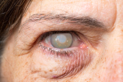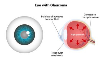
Gonioscopy is a specialised eye examination used to assess the internal drainage system of the eye. It allows ophthalmologists to observe the angle where the iris meets the cornea, which is essential for the proper outflow of aqueous fluid. This test plays a vital role in diagnosing glaucoma, a condition that can lead to irreversible vision loss if untreated.
By providing detailed insights into the eye’s drainage structure, gonioscopy helps doctors determine the type and severity of glaucoma and plan appropriate management strategies. It is a quick, painless, and highly effective test for maintaining long-term eye health.
What is Gonioscopy?
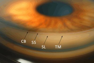
Gonioscopy is a diagnostic eye test that enables doctors to view the anterior chamber angle, the area responsible for fluid drainage in the eye. The gonioscopy test helps identify whether the drainage angle is open, narrow, or closed, which are key indicators for detecting glaucoma and related eye disorders.
A special lens called a gonioscope is placed on the eye’s surface to visualise this area clearly. This test is an essential part of a comprehensive eye examination and is typically recommended when there are signs of elevated eye pressure or suspicion of angle-closure glaucoma.
When is the Gonioscopy Test Performed?
Doctors recommend a gonioscopy test when there are signs that the eye’s drainage system may be compromised. This may include high intraocular pressure (IOP), a family history of glaucoma, or abnormalities observed during a routine eye exam.
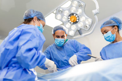
It is also performed before certain surgeries, after eye trauma, or in patients with ocular hypertension. Regular gonioscopy examinations are crucial for individuals at risk of angle-closure glaucoma, as early detection enables preventive intervention and helps safeguard long-term vision.
Gonioscopy Test Procedure
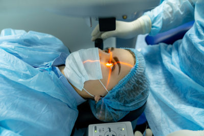
The gonioscopy test procedure is simple, safe, and takes only a few minutes. First, the eye is numbed using anaesthetic eye drops to ensure comfort. The ophthalmologist then gently places a gonioscopy lens on the cornea. Using a slit-lamp biomicroscope, the doctor examines the angle formed between the iris and cornea.
The reflections from the lens’s mirrored surfaces allow the doctor to assess the drainage angle’s structure, helping detect blockages or abnormal formations. The procedure is painless and does not affect vision after completion.
How Does the Gonioscopy Test Work?
The gonioscopy test provides a clear view of the anterior chamber angle, an area not visible during routine examination because of total internal reflection within the cornea. The gonioscope lens neutralises this reflection, allowing the ophthalmologist to observe the drainage channels directly.
Through this process, doctors can identify abnormalities such as angle recession, neovascularisation, or pigment dispersion, which can lead to glaucoma. The test’s accuracy in assessing the eye’s fluid outflow pathway makes it an essential diagnostic tool for glaucoma management.
Types of Gonioscopy Lenses
There are two main types of gonioscopy techniques used in clinical practice – direct and indirect gonioscopy. The choice depends on the type of examination required and the comfort of both the patient and the doctor.
Direct Gonioscopy
Direct gonioscopy uses a Koeppe lens to provide a direct view of the anterior chamber angle. This technique is typically performed with the patient lying down under a microscope.
The Koeppe lens provides a detailed, magnified view of the angle structures, making it useful for surgical or research purposes. Although it provides high clarity, direct gonioscopy is less commonly used in routine clinical practice.
Indirect Gonioscopy
Indirect gonioscopy uses lenses such as the Goldmann or Zeiss gonioscopy lens along with a slit-lamp biomicroscope. These lenses contain mirrors that allow the doctor to view the drainage angle from different perspectives.
It is more practical for clinical use and provides a wide, panoramic view of the anterior chamber. Because of its efficiency and comfort, indirect gonioscopy is the preferred method in most eye clinics.
Gonioscopy Test Benefits and Uses in Eye Health
The gonioscopy test plays a crucial role in the early detection and management of glaucoma. It helps determine whether the eye has open-angle or angle-closure glaucoma, guiding treatment.
In addition, gonioscopy is valuable for diagnosing conditions such as pigmentary glaucoma, neovascular glaucoma, and angle recession caused by trauma. By assessing the angle structures, doctors can identify obstructions that impede fluid drainage and elevate intraocular pressure. The test ensures accurate diagnosis, timely intervention, and improved visual outcomes.
What to Expect During Gonioscopy Test
The gonioscopy test is painless, quick, and non-invasive. Before the test, the doctor applies anaesthetic drops to prevent discomfort. During the examination, the patient rests their chin on the slit-lamp support while the doctor gently places the gonioscopy lens on the eye.
The lens does not cause pain or pressure. Patients may see a bright light or mild reflections but experience no lasting discomfort. After the test, normal activities can be resumed immediately, and vision remains unaffected.
Results and Follow-Up After Gonioscopy Test
After a gonioscopy test, the doctor reviews the findings to determine the structure and condition of the drainage angle. If abnormalities are detected, such as narrow angles, angle closure, or scarring, the ophthalmologist may recommend further diagnostic tests or initiate treatment.
For patients with normal findings, regular monitoring may still be advised to track changes over time. In cases of glaucoma, follow-up appointments include IOP checks, visual field testing, and optic nerve imaging to assess disease progression.
When to Visit a Doctor for a Gonioscopy Test?
It is recommended to undergo a gonioscopy test if you experience eye pain, halos around lights, blurred vision, or a sudden change in visual clarity. People with a family history of glaucoma, eye trauma, or high intraocular pressure should schedule periodic gonioscopy evaluations.
Early testing is crucial for those above 40 years of age or with risk factors like diabetes and hypertension, as these conditions increase the likelihood of developing glaucoma.