The eyes are used to create a complete and balanced image in vision. When this harmony is broken and one eye sees objects bigger or smaller than the other, it is known as aniseikonia.
This visual distortion is not very common, but it may severely influence the sense of depth, coordination and comfort. Understanding the causes, signs, and remedies of aniseikonia helps patients and clinicians manage the problem effectively and enhance visual performance.
What Is Aniseikonia?
Aniseikonia is a condition in which there is an apparent difference in size or shape between the images from the two eyes. This mismatch prevents proper image fusion, leading to visual incongruence, headaches, eye strain, and, in more serious cases, balance loss.
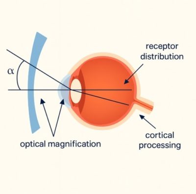
Our two eyes usually record similar images, which the brain combines to form a single three-dimensional picture. However, when one image appears larger or smaller, the brain struggles to align them accurately, disrupting binocular vision. This condition can be optical, retinal, or cortical in origin and may develop after eye surgery, refractive errors, or retinal diseases.
Types of Aniseikonia
Optical
This form occurs when an image size disparity is due to optical factors, e.g., large variations in refractive errors (anisometropia) or improperly corrected lenses. For example, if one lens has greater magnifying power than the other, the two eyes will perceive the image differently. Specialised lenses, or contact lenses, are used to correct optical aniseikonia and equalise magnification.
Retinal
Retinal aniseikonia occurs when there are variations in the structure or function of the retinas of the two eyes.
Other conditions, like macular oedema, retinal detachment, or epiretinal membrane, may cause uneven stretching or swelling of the retina, leading to unequal image scaling. Medical or surgical treatment of the underlying retinal pathology may be necessary before any attempt to improve visual correction is made.
Cortical
This type is caused by how the brain processes visual information from both eyes. Even when the optical and retinal images are identical, neurological factors or brain injuries can disrupt image interpretation, leading to perceived size differences. Cortical aniseikonia is less common but may occur following trauma, stroke, or neurological disease affecting the visual cortex.
What Causes Aniseikonia?
Common causes include:
- Refractive Differences (Anisometropia): When the prescription of one eye differs from the other, it may result in unequal magnification of images.
- Cataract Surgery: In each eye, the insertion of various intraocular lenses (IOLs) is liable to cause an imbalance of image sizes.
- Refractive Surgery (LASIK or PRK): The eyes can have their images magnified or distorted by altering the cornea’s shape.
- Retinal Diseases: Macular oedema, retinal detachment, or epiretinal membranes distort the structure of the retina and the size of the image.
- Trauma or Neurological Disorders: The brain damage may interfere with the interpretation of visual information.
- Poor Eyewear: Lenses that are not fitted properly or new types of lenses may increase the amount of perceived size variation.
Common Symptoms of Aniseikonia
Patients often report one or more of the following:
- Double Vision or Blurred Vision: The inability to unite images because of their dissimilar sizes.
- Eye Strain and Headaches: These are caused by the continuous struggle to organise and integrate pictures.
- Dizziness or Balance Problems: Interruption in space perception and depth.
- Skewed or Lopsided Vision: Objects can be distorted, stretched, or have different sizes.
- Difficulties in Reading: My fatigue, lines skipped or discomfort with long reading.
Diagnosis of Aniseikonia
Diagnosis of aniseikonia encompasses clinical examination, as well as special visual examination.
- Subjective Tests: The New Aniseikonia Test/space eikonometer measures the percentage change in image perception by the eyes.
- Objective Evaluation: Retinal imaging, optical coherence tomography (OCT) and refraction test are used to determine the cause, including underlying retinal diseases or optical distortions.
- Evaluation of Binocular Vision: Alignment of the eyes and the ability to fuse are assessed to determine how the brain combines images.
Treatment Options for Aniseikonia
Optical Corrections (Glasses or Contact Lenses)
The most widespread treatment of aniseikonia is specialised optical solutions. Special-purpose lenses, such as iseikonic lenses, are custom-made lenses that adjust magnification to match the sizes of images seen by the two eyes. Anisometropia differences can also be minimised using contact lenses.
Vision Therapy and Rehabilitation
Vision therapy can train the eyes and brain to adapt to image differences and restore better binocular coordination. Exercises may include fusion training, vergence control, and improvement of depth perception. When adaptation is possible, gradual adjustment with optical aids and vision therapy offers long-term comfort and stability.
Surgical Options in Complex Cases
When aniseikonia results from structural or retinal abnormalities, surgical intervention may be necessary. For example:
- Retinal surgery for detachment or macular disorders can restore symmetry.
- Refractive correction or IOL replacement may balance image magnification after cataract surgery.
Living with Aniseikonia
Living with aniseikonia can be challenging, particularly when symptoms cause discomfort or interfere with reading, driving, or coordination. However, several coping strategies can help manage the condition effectively:
- Regular Eye Examinations: Ongoing monitoring ensures timely adjustments in prescriptions or therapy.
- Proper Eyewear Fitting: Customised glasses or contact lenses reduce distortion and strain.
- Gradual Adaptation: The brain may take time to adjust to new lenses or corrective treatments.
- Lifestyle Adjustments: Adequate lighting and regular breaks during visual tasks reduce eye strain.
Conclusion
Aniseikonia is a vision condition where one eye perceives images differently in size or shape from the other, leading to discomfort and visual imbalance. Understanding the meaning of aniseikonia, identifying its causes, and undergoing appropriate tests are key to accurate diagnosis.
With the right combination of optical correction, therapy, and medical care, most patients can regain comfortable binocular vision. Regular eye check-ups, adherence to prescribed treatment, and collaboration with an experienced eye specialist ensure better long-term outcomes and quality of life.