Retinal changes often develop silently, without obvious warning signs. One such finding is dot and blot hemorrhages, a common yet important sign of underlying retinal or systemic disease.
Although patients cannot see these hemorrhages themselves, they are frequently detected during routine eye examinations and can provide early clues about conditions such as diabetes or hypertension. This blog explains what dot and blohemorrhages are, why they occur, how they affect vision, and how they are managed.
Understanding Dot and Blot hemorrhages
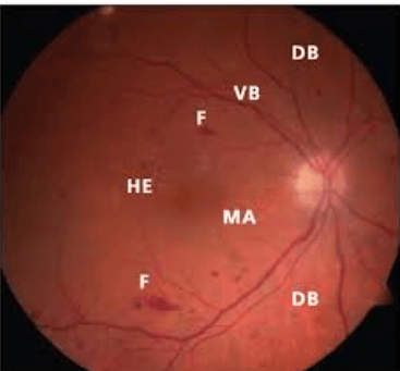
Dot and blot hemorrhages are small intraretinal bleeds that occur in the deeper layers of the retina, specifically the inner nuclear and outer plexiform layers. They appear as round or irregular dark red spots when the retina is examined through dilated pupils.
Because these hemorrhages form deep within the retina, they do not cause visible redness, pain, or irritation of the eye. Instead, they are usually discovered incidentally during eye screening or evaluation for blurred vision. Clinically, dot and blot retinal hemorrhages often appear together and are an important marker of retinal vascular damage.
Dot vs. Blot – What’s the Difference?
|
Feature |
Dot Hemorrhages |
Blot Hemorrhages |
|
Appearance |
Tiny, round red spots. |
Slightly larger, “blob-shaped” spots. |
|
Origin |
Caused by leakage from tiny, weakened capillaries. |
Arise when blood spreads more widely within the retinal tissue. |
|
Location |
Seen in the deeper layers of the retina. |
Distributed across multiple retinal layers. |
|
Significance |
Indicates compromised vessel integrity. |
Indicates more extensive vascular leakage. |
What Causes Dot and Blot Hemorrhages?
The causes of dot and blot hemorrhages are linked to damage or stress affecting the retinal microvasculature. The most common causes include:
- Diabetic retinopathy: Chronic high blood sugar damages retinal capillaries, making them leaky and prone to rupture.
- Hypertensive retinopathy: Persistently high blood pressure weakens vessel walls.
- Retinal vein occlusion: Blockage of retinal veins increases venous pressure, leading to capillary rupture.
- Blood disorders: Severe anaemia, clotting abnormalities, and autoimmune conditions can increase the risk of bleeding.
- Medications and trauma: Anticoagulants, trauma, or birth-related injury may also contribute.
In clinical practice, dot and blot hemorrhages seen in diabetic retinopathy and retinal vein occlusion are particularly significant, as they may indicate disease progression or poor systemic control.
Recognising Symptoms of Dot and Blot Hemorrhages
Many people with dot and blothemorrhages retinopathy do not experience symptoms in the early stages. When symptoms occur, they may include:
- Mild blurring or distortion of vision
- Small dark spots or floaters
- Difficulty reading fine print
- Reduced contrast sensitivity
- Problems with night vision in advanced cases
Symptoms often depend on whether the macula is involved. Sudden or noticeable changes in vision should always prompt an urgent eye examination.
Diagnosis – How Are Dot and Blot Hemorrhages Detected?
Diagnosis of dot and blotheamorrhages relies on a detailed retinal assessment performed by an eye specialist. Key diagnostic tools include:
- Dilated fundus examination to visualise the retina directly
- Optical coherence tomography (OCT) to assess retinal layers and detect macular oedema
- Fluorescein angiography to identify leaking or blocked blood vessels
- Systemic evaluation, including blood sugar levels, blood pressure, and blood tests
Identifying the underlying systemic cause is essential, as treating the retinal findings alone is not sufficient.
Treatment Options for Dot and Blot Hemorrhages
Dot and blot hemorrhage treatment focuses primarily on addressing the underlying cause rather than the hemorrhage itself.
Managing the Underlying Cause
- Tight blood sugar control in diabetes reduces further retinal damage
- Blood pressure control protects fragile retinal vessels
- Treatment of anaemia, clotting disorders, or autoimmune disease when present
- Review of blood-thinning medications under medical supervision
Manhemorrhageses gradually resolve once systemic control improves.
Medical and Laser Therapies
Hemorrhages are associated with macular oedema, abnormal vessel growth, or vision loss:
- Laser photocoagulation may be used to seal leaking vessels
- Anti-VEGF injections help reduce retinal swelling and vascular leakage
- Vitrectomy may be required for severe complications such as vitreous hemorrhage
Treatment decisions are individualised based on severity and retinal findings.
Lifestyle and Home Care
Lifestyle changes support retinal health and include:
- A balanced diet and regular physical activity
- Smoking cessation
- Adherence to medical treatment plans
- Regular eye examinations, especially for people with diabetes or hypertension
Early detection through routine screening remains one of the most effective strategies.
Emerging Research and Future Treatments
Advances in retinal imaging and artificial intelligence are improving the early detection of dot and blot hemorrhages. Sustained-release therapies and improved screening programmes aim to reduce treatment burden and preserve vision more effectively.
Preventing Dot and Blot Hemorrhages
Preventing dot and blot hemorrhages involves long-term control of systemic risk factors:
- Maintain stable blood sugar and blood pressure
- Manage cholesterol and cardiovascular health
- Attend routine dilated eye examinations
- Follow prescribed medications consistently
Proactive care significantly reduces the risk of progression to sight-threatening disease.
Living with Dot and Blot Hemorrhages
Most individuals retain good vision when these hemorrhages are detected early, and underlying conditions are well controlled. Ongoing monitoring is essential, as recurrence may signal worsening systemic disease. With appropriate care, long-term visual outcomes are often favourable.
Conclusion – Protecting Your Vision from Dot and Blot Hemorrhages
Dot and blothemorrhages are important retinal signs that reflect deeper vascular health issues. While often asymptomatic initially, they can indicate progressive conditions such as diabetes or hypertension. Early diagnosis, systemic control, and timely ophthalmic treatment help preserve vision. Regular eye examinations remain the cornerstone of prevention and long-term eye health.