
Optic atrophy, also called optic nerve atrophy, is damage to the optic nerve — the cable that carries visual information from the eye to the brain. When this nerve is damaged, the signals it sends weaken or stop, leading to vision loss that is often permanent. Optic atrophy is not a single disease but the end result of many possible conditions affecting the optic nerve. In this guide, we explain what optic atrophy means, its causes, symptoms, types, and the treatment options available — and what you can realistically expect.
Optic Atrophy Meaning and How It Affects Vision
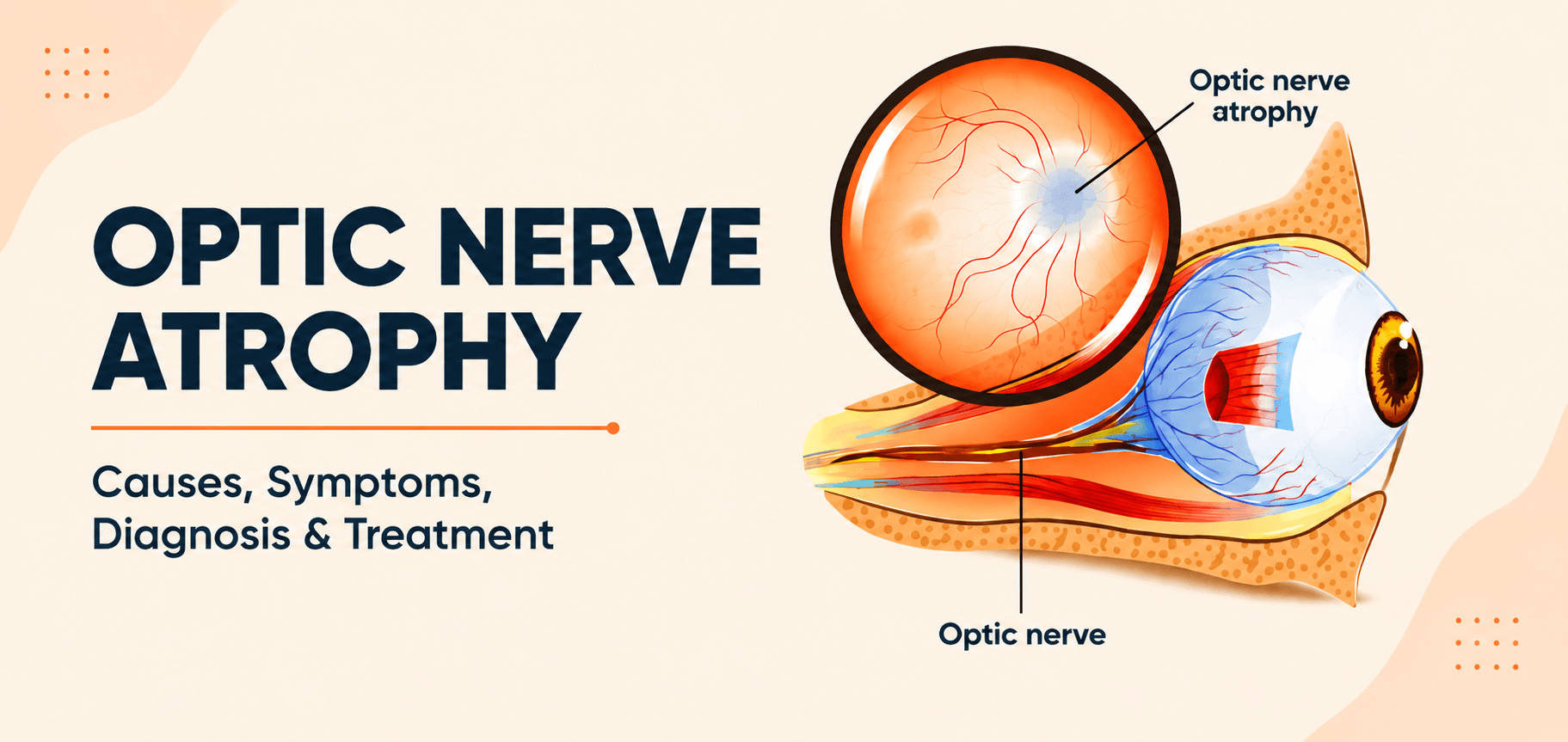
The meaning of optic atrophy is straightforward: “atrophy” means wasting or shrinkage, so optic atrophy refers to the degeneration of the optic nerve fibres. As these fibres are damaged, the nerve can no longer transmit visual signals from the retina to the brain effectively, and vision is lost in the affected eye.
An important point to understand early is that optic nerve damage is usually permanent, because the nerve fibres do not regenerate. However, the situation is not always hopeless — if the underlying cause is treated in time, further damage can often be prevented and remaining vision preserved.
How the Optic Nerve Works in Normal Vision
The optic nerve is made up of more than a million tiny nerve fibres. When light enters the eye, the retina converts it into electrical signals. The optic nerve carries these signals from the retina to the brain, which interprets them as the images you see. In healthy vision, this transmission is fast and seamless — the optic nerve is the essential link between the eye and the brain.
What Happens When Optic Atrophy Develops
When optic atrophy develops, the nerve fibres progressively degenerate. As more fibres are lost, fewer signals reach the brain, and vision deteriorates. On examination, the optic disc (the visible head of the optic nerve at the back of the eye) appears pale instead of its normal healthy pink. Because the damage reflects nerve fibre loss, the resulting vision loss is typically irreversible — which is why recognising the signs of optic nerve damage early is so important.
What Causes Optic Atrophy? Key Risk Factors Explained
There are many optic atrophy causes, ranging from circulation problems to inherited conditions. The main causes include:
Reduced Blood Supply to the Optic Nerve
When blood flow to the optic nerve is interrupted — a condition called ischemic optic neuropathy — the nerve is starved of oxygen and can be permanently damaged. This is a common cause, particularly in older adults and those with vascular risk factors.
Infections and Inflammation
Inflammation of the optic nerve (optic neuritis) and infections such as meningitis can damage nerve fibres. Optic neuritis is sometimes linked to neurological conditions like multiple sclerosis.
Genetic and Hereditary Conditions
Some forms of optic atrophy are inherited. Leber hereditary optic neuropathy (LHON) and dominant optic atrophy are genetic conditions that damage the optic nerve, often appearing in childhood or young adulthood.
Trauma or Injury to the Eye or Brain
A direct injury to the eye, or a head injury affecting the optic nerve or its pathway, can lead to optic nerve damage and subsequent atrophy.
Brain Tumors or Neurological Disorders
Tumours or other lesions that press on the optic nerve or its pathway in the brain can compress the nerve, gradually causing atrophy. This is why unexplained vision loss sometimes requires brain imaging.
Long-Term Eye Conditions Like Glaucoma
Chronic eye diseases can also be responsible. In particular, untreated [glaucoma — internal link] raises pressure inside the eye and slowly damages the optic nerve, leading to glaucomatous optic atrophy.
Common Optic Atrophy Symptoms You Should Not Ignore
Optic atrophy symptoms depend on the cause and how advanced the damage is, but they generally involve a noticeable decline in vision. Watch for these signs of optic nerve damage:
Gradual or Sudden Vision Loss
Vision may fade slowly over time or, in some causes such as ischemia, drop suddenly. Any unexplained vision loss needs prompt assessment.
Reduced Color Vision
Colours may appear washed out or less vivid, with red in particular looking dull — an early and characteristic sign of optic nerve problems.
Difficulty Seeing Details (Blurred Vision)
Reduced sharpness and difficulty making out fine detail are common, as the nerve can no longer relay clear, detailed information to the brain.
Loss of Peripheral Vision (Tunnel Vision)
Some people lose their side vision, leaving a narrowed field of sight — often described as tunnel vision.
Abnormal Pupil Reaction
When one eye is more affected than the other, the pupil may respond abnormally to light (a relative afferent pupillary defect), which a doctor can detect during an examination.
Optic Atrophy Types and Classification Explained
Doctors classify optic atrophy by how the damage occurred. The main optic atrophy types are:
Primary Optic Atrophy
Here the optic nerve is damaged directly, without prior swelling of the disc. The optic disc looks pale with clearly defined, sharp margins. Causes include compression, hereditary conditions, and certain neuropathies.
Secondary Optic Atrophy
This follows previous swelling or inflammation of the optic disc (such as papilloedema or papillitis). The pale disc has blurred, indistinct margins because of the earlier swelling.
Consecutive Optic Atrophy
This type results from diseases of the retina — such as retinitis pigmentosa or a retinal artery occlusion — that damage the nerve fibres feeding into the optic nerve.
Glaucomatous Optic Atrophy
Caused by glaucoma, this form is marked by characteristic “cupping” of the optic disc due to long-standing raised eye pressure.
Primary vs Secondary Optic Atrophy: Key Differences
|
Feature |
Primary Optic Atrophy |
Secondary Optic Atrophy |
|
Preceding disc swelling |
No |
Yes (follows swelling/inflammation) |
|
Optic disc margins |
Sharp, well defined |
Blurred, indistinct |
|
Disc appearance |
Pale, clean |
Pale with surrounding changes |
|
Typical causes |
Compression, hereditary, retrobulbar neuritis |
Papilloedema, papillitis |
How Doctors Diagnose Optic Atrophy
Diagnosing optic atrophy combines a clinical eye examination with imaging to confirm nerve damage and find its cause.
Eye Examination (Fundoscopy)
The doctor examines the optic disc with an ophthalmoscope, looking for the tell-tale pallor of optic atrophy.
Visual Field Test
A perimetry test maps your field of vision to detect blind spots or loss of peripheral vision caused by nerve damage.
OCT Scan (Optical Coherence Tomography)
An OCT scan measures the thickness of the retinal nerve fibre layer, objectively confirming and quantifying optic nerve fibre loss.
MRI or Brain Imaging
When a compressive, inflammatory, or neurological cause is suspected, an MRI of the brain and orbits helps identify tumours, inflammation, or other problems along the optic pathway.
Optic Atrophy Treatment Options and Management
There is no treatment that regrows a damaged optic nerve, so optic atrophy treatment focuses on stopping further damage and making the most of remaining vision.
Treating the Underlying Cause
The most important step is addressing the cause — for example, treating an infection or inflammation, relieving pressure from a tumour, or controlling eye pressure in glaucoma. This can halt progression and protect the vision that remains.
Medications and Neuroprotective Support
Depending on the cause, medications such as corticosteroids (for inflammatory conditions) may be used. Neuroprotective approaches are an area of ongoing research and are used selectively under specialist guidance.
Vision Rehabilitation and Low Vision Aids
For those living with reduced vision, low-vision aids such as magnifiers and specialised devices, along with vision rehabilitation services, can significantly improve day-to-day independence.
Lifestyle Changes to Protect Remaining Vision
Managing general health — controlling blood pressure and diabetes, eating well, avoiding smoking and excess alcohol, and protecting the eyes — all help safeguard the remaining optic nerve fibres.
Is There an Optic Atrophy Cure? What Patients Should Know
Many patients ask whether there is an optic atrophy cure. It is important to have realistic expectations.
Can Optic Nerve Damage Be Reversed?
At present, established optic nerve damage cannot be reversed, because the nerve fibres do not regenerate. Treatment cannot restore vision that has already been lost to atrophy.
When Early Treatment Can Help Preserve Vision
The encouraging part is timing. If the underlying cause is identified and treated before the nerve is fully damaged, further loss can often be prevented, and in some cases vision can be stabilised or partially preserved. This is why early diagnosis makes such a difference.
How to Prevent Optic Atrophy or Reduce Risk
While not every cause is preventable, you can lower your risk and protect your optic nerve:
Regular Eye Check-ups
Routine [comprehensive eye check-ups — internal link] can detect optic nerve problems and conditions like glaucoma early, before significant damage occurs.
Managing Chronic Conditions Like Diabetes and BP
Keeping diabetes and blood pressure well controlled protects the small blood vessels that supply the optic nerve.
Protecting Eyes from Injury
Using protective eyewear during risky activities helps prevent the kind of trauma that can damage the optic nerve.
Early Treatment of Eye Diseases
Promptly treating eye diseases — especially glaucoma and any [retinal diseases — internal link] — prevents the slow nerve damage that leads to atrophy.
When Should You See an Eye Specialist?
Seek prompt assessment by an eye specialist if you notice any of the following warning signs:
- Sudden loss of vision in one or both eyes
- A change in colour vision, such as colours looking faded
- Loss of side (peripheral) vision
These symptoms can signal optic nerve damage that needs urgent evaluation, as early action offers the best chance of protecting your sight.
Conclusion
Optic atrophy reflects permanent damage to the optic nerve and is an important cause of vision loss, but understanding it helps you act in time. While the condition may not always be reversible, identifying and treating the underlying cause early can halt its progression and preserve valuable remaining vision. If you experience any sudden vision change, colour vision loss, or unexplained visual decline, consult an eye specialist without delay — early diagnosis is the key to protecting your sight.