


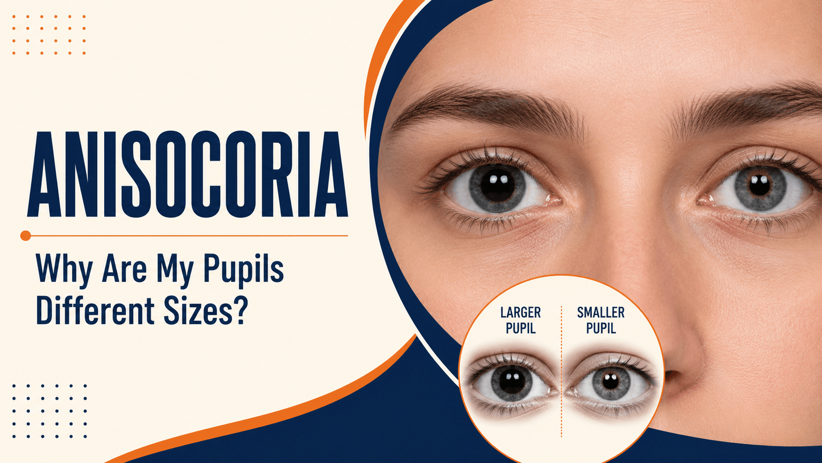
You glance in the mirror, or look closely at a photograph, and notice that one pupil looks larger than the other. It is an unsettling thing to spot, and the first question is always the same: does this matter?
For a large number of people, the answer is no. Unequal pupils are a normal variation in a substantial minority of healthy adults. But a small number of causes are serious and time-sensitive, so it is worth knowing which is which.
Key Takeaway: What Is Anisocoria?
Anisocoria is the medical term for pupils of unequal size. It is common and often completely harmless; a benign variant called physiological anisocoria affects roughly one in five people. However, anisocoria that appears suddenly, or comes with a droopy eyelid, double vision, eye pain, severe headache or neck pain, needs urgent medical assessment, because a few causes are serious.
What Is Anisocoria?
The pupil is the black opening at the centre of the coloured iris. It widens in dim light to let more light in, and narrows in bright light to let less in. Two sets of nerves control this: one set constricts the pupil, the other dilates it.
Anisocoria simply means the two pupils are different sizes. It is a sign, not a diagnosis. The important question is never the size difference itself, but what is causing it and whether the problem lies in the eye, in the nerves supplying it, or in the medicines a person has been exposed to.
How Common Are Unequal Pupils?
More common than most people expect. Physiological anisocoria, a harmless, lifelong difference in pupil size with no underlying disease, is estimated to affect around 20% of the population, although some more recent measurements suggest a somewhat lower figure.
In this benign form, the difference is usually small, typically under about one millimetre, and it stays roughly the same whether the room is bright or dark. Both pupils still react normally to light. Many people have it for years without ever noticing.
Which Pupil Is the Abnormal One? The Light-and-Dark Clue
This is the most useful idea in the whole topic, and it is what an eye doctor works out first.
A pupil should constrict in bright light and dilate in darkness. So if you compare the two pupils in both conditions, the pattern reveals which one is failing to do its job.
|
What you observe |
Which pupil is abnormal |
What it suggests |
|
Difference is greater in dim light or darkness |
The smaller pupil is not dilating properly |
Horner syndrome, or physiological anisocoria |
|
Difference is greater in bright light |
The larger pupil it is not constricting properly |
Third nerve palsy, Adie tonic pupil, dilating medication, iris injury |
|
Difference stays about the same in light and dark, and is small |
Usually neither |
Most likely physiological anisocoria |
A word of caution: this is a way of understanding what your doctor is assessing, and it gives you something useful to describe at your appointment. It is not a way to diagnose yourself. Several causes overlap in appearance, and a proper examination is needed to separate them.
Common Causes of Anisocoria
Physiological anisocoria
The most common cause by far. The size difference is small, present in both light and dark, and unaccompanied by any other symptom. Vision, eye movements and eyelid position are all normal. No treatment is needed.
Horner syndrome
Here the affected pupil is smaller than normal, because the nerve supply that dilates it has been interrupted somewhere along its path. The classic combination is a small pupil, a mildly droopy eyelid (ptosis) and reduced sweating on the same side of the face, though not every feature is always present.
Horner syndrome matters because of what can cause it. The nerve pathway travels a long route through the neck and chest, so causes include a tear in the carotid artery in the neck (carotid dissection), a tumour at the top of the lung, thyroid or neck masses, and problems within the brain or spinal cord. New Horner syndrome, particularly with neck or face pain, needs urgent assessment.
Third nerve palsy
Here the affected pupil is larger and reacts poorly to light, because the nerve that constricts it is not working. It is usually accompanied by a drooping eyelid, and often by double vision and difficulty moving the eye in certain directions, a pattern sometimes described as a paralytic squint.
A new third nerve palsy involving the pupil is treated as a medical emergency, because one recognised cause is an aneurysm pressing on the nerve. Anyone who develops a dilated pupil together with a droopy eyelid and double vision, especially with headache, should go to an emergency department immediately for brain imaging, not wait for an outpatient appointment.
Adie tonic pupil
In this condition the affected pupil is larger, responds sluggishly or not at all to light, and constricts slowly when focusing on something close. It is generally benign and most often affects younger adults, more commonly women. Some people notice glare or difficulty focusing for near work. It still needs proper evaluation to exclude other causes, but it is not dangerous in itself.
Medicines and eye drops
Pharmacological causes are common and easily missed. A pupil can become dilated after accidental contact with dilating drops used during an eye examination, or from medication residue transferred by the fingers. Nebulised medication escaping around a mask can dilate the pupil on one side. Motion-sickness patches containing scopolamine are another recognised source. Conversely, some glaucoma drops constrict the pupil, so treatment in one eye only can produce a visible difference.
A helpful clue: if the eye with the larger pupil also has noticeably blurred near vision, a dilating medication is a likely explanation.
Eye injury
Blunt trauma can damage the small muscle that constricts the pupil, leaving it permanently larger and irregular in shape. Any anisocoria that follows an eye or head injury should be examined promptly.
Inflammation and previous eye surgery
Inflammation inside the eye, such as uveitis or iritis, can make a pupil smaller or irregular and is usually accompanied by pain, redness and light sensitivity. Previous eye surgery can also leave a pupil slightly altered in size or shape, which is expected rather than concerning.
Causes at a Glance
|
Cause |
Affected pupil |
Typical accompanying signs |
Urgency |
|
Physiological anisocoria |
Neither; small difference |
None |
Not urgent |
|
Horner syndrome |
Smaller |
Droopy lid, reduced facial sweating, sometimes neck pain |
Urgent if new |
|
Third nerve palsy |
Larger |
Droopy lid, double vision, limited eye movement |
Emergency |
|
Adie tonic pupil |
Larger |
Poor light reaction, slow near focusing, glare |
Needs assessment; usually benign |
|
Medication or drops |
Usually larger |
Blurred near vision in that eye; recent exposure |
Needs assessment |
|
Eye injury |
Usually larger, irregular |
History of trauma |
Prompt assessment |
|
Uveitis or iritis |
Often smaller, irregular |
Pain, redness, light sensitivity |
Prompt assessment |
When Unequal Pupils Are a Medical Emergency
Seek emergency medical care immediately if unequal pupils appear suddenly, or occur with any of the following:
- A drooping eyelid together with a dilated pupil, especially with double vision
- Severe or unusual headache
- Neck pain or face pain alongside a small pupil and droopy lid
- Eye pain, redness or loss of vision
- Weakness, numbness, slurred speech, confusion or difficulty walking
- Recent head, eye or neck injury
- Fever with neck stiffness
These patterns can reflect a problem in the nerves or blood vessels rather than the eye itself, and the causes involved are treatable when identified early.
Other Symptoms to Notice Alongside Anisocoria
Even when nothing feels urgent, these details help your doctor and are worth noting before your appointment:
- Whether the difference is more obvious in a bright room or a dark one
- Whether one eyelid sits lower than the other
- Any double vision, blurring or difficulty focusing up close
- Glare or discomfort in bright light
- Recent illness, injury, surgery, or new medicines and eye drops
- Whether the difference comes and goes, or is constant
How Anisocoria Is Diagnosed
Assessment is painless and follows a logical sequence.
- History. When it was first noticed, whether it came on suddenly, any injury, illness, surgery or medication exposure.
- Pupil examination in light and darkness. Measuring both pupils in each condition to establish which one is behaving abnormally.
- Pupil reaction testing. Checking how each pupil responds to light, and how it behaves when focusing on a near object.
- Eyelid and eye movement assessment. Looking for ptosis and for restricted eye movements, which help distinguish third nerve palsy from Horner syndrome.
- Slit-lamp examination. Inspecting the iris for injury, inflammation or structural change, and checking the front of the eye.
- Pharmacological pupil testing where needed. Specific diagnostic eye drops can confirm conditions such as Horner syndrome or an Adie pupil.
- Imaging and further tests when indicated. MRI, CT or vascular imaging of the head and neck if a nerve or blood vessel cause is suspected.
Because the possible causes range from the eye to the neck, chest and brain, assessment sometimes involves a neuro-ophthalmology opinion or joint care with a neurologist or physician.
How Anisocoria Is Treated
There is no treatment for anisocoria itself, because it is a sign rather than a disease. Treatment is directed entirely at the underlying cause.
- Physiological anisocoria needs no treatment at all, only reassurance.
- Medication-related dilation resolves on its own once the exposure stops, usually within hours to a few days depending on the substance.
- Horner syndrome and third nerve palsy are managed by treating the underlying condition, which may involve urgent medical or surgical care.
- Adie tonic pupil may need no treatment. Where glare or near-focusing difficulty is troublesome, tinted lenses or reading correction can help.
- Inflammation is treated with prescribed anti-inflammatory eye medication under specialist supervision.
- Traumatic iris damage may be left alone if vision is comfortable, or managed with tinted lenses or, in selected cases, surgical repair.
Outcomes depend on the cause, and no single approach suits every patient.
What to Do If You Notice Unequal Pupils
- Check for emergency features first: droopy lid, double vision, severe headache, neck pain, eye pain, weakness, or slurred speech. If any are present, seek emergency care now.
- Look at old photographs. This is genuinely useful. If the same difference is visible in pictures from years ago, the anisocoria is long-standing and far more likely to be benign. If earlier photos show equal pupils, it is new and needs assessment.
- Note the pattern. Observe whether the difference is greater in a bright or dim room, and take a clear photograph in each condition to show your doctor.
- Review recent exposures. New eye drops, patches, nebulisers, or handling of any medication.
- Book an eye examination. Even reassuring-looking anisocoria deserves confirmation, and a comprehensive eye examination can establish the cause.
Do not use any eye drops to try to even out the pupils. Doing so masks the picture and makes accurate diagnosis harder.
Anisocoria in Children
Unequal pupils in children follow the same principles, with a few additions. Physiological anisocoria occurs in children too and is harmless.
Two situations warrant particular attention. A small pupil with a droopy eyelid present from birth or early infancy, sometimes with a difference in iris colour between the eyes, should be assessed, as congenital Horner syndrome needs evaluation. And any newly noticed anisocoria in a child, especially after a fall or head injury, should be examined promptly rather than watched at home.
Parents often spot this in photographs before anyone else does. Bringing those photos to the appointment genuinely helps.
Conclusion
Anisocoria is common, and for a large number of people it is simply how their eyes have always been. The reassuring pattern is a small, long-standing difference with no other symptoms and normal vision, often confirmed by nothing more sophisticated than an old photograph.
What changes the picture is newness and company. Unequal pupils that appear suddenly, or arrive alongside a droopy eyelid, double vision, headache, neck pain or eye pain, deserve immediate medical attention rather than observation.
If you have noticed a difference in your pupils and are unsure which situation applies, have it checked properly. Book an appointment with an eye specialist, and bring older photographs with you; they are often the most helpful thing you can provide.