



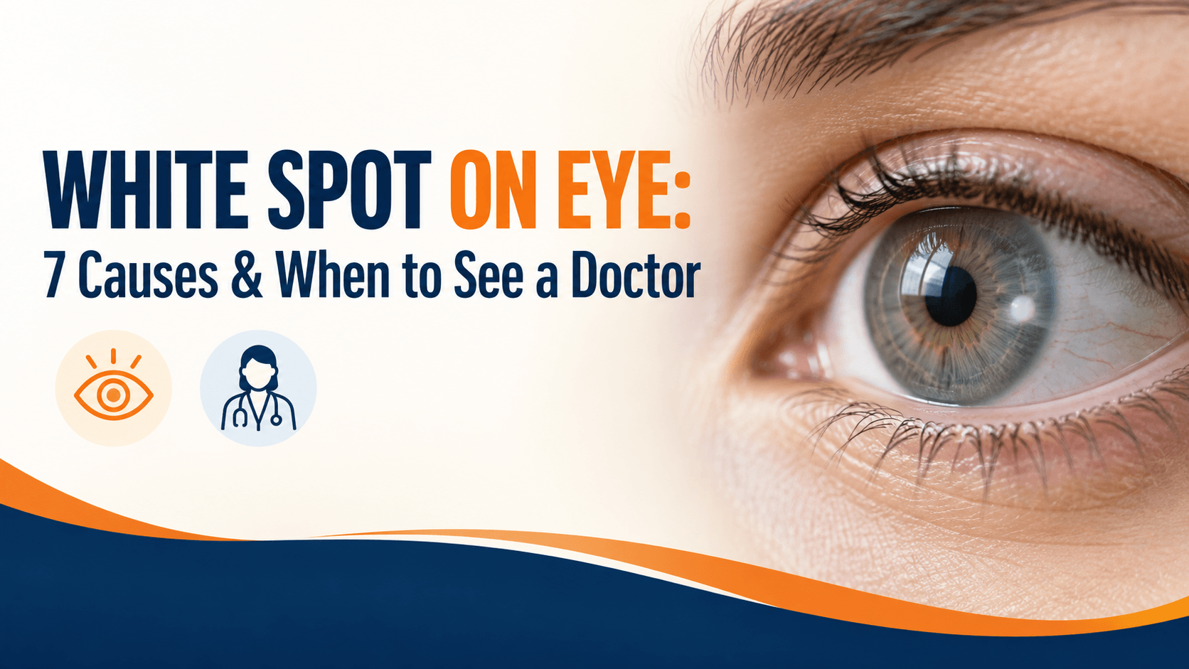
Most white spots on the eye turn out to be harmless. A few are emergencies. The difference is usually not how the spot looks; it is where it sits.
Key Takeaway
A white spot on the white part of the eye is usually a harmless growth such as a pinguecula. A white spot on the clear cornea, especially with pain, redness or blurred vision, may be a corneal ulcer and needs same-day care. A white reflex inside a child’s pupil needs urgent eye examination, because it can indicate a serious condition of the retina.
First, Where Exactly Is the White Spot?
Before reading about causes, look in a mirror in good light and work out which part of the eye the spot is on. This one observation sorts most cases.
|
Where the spot is |
What it usually means |
How soon to see a doctor |
|
On the white of the eye (sclera/conjunctiva) |
Usually a harmless growth or deposit |
Routine appointment |
|
On the clear cornea, over the iris or pupil, with pain or redness |
Possible infection or ulcer |
Same day |
|
On the clear cornea, no pain, longstanding |
Often an old scar |
Within a few weeks |
|
Inside the pupil, a white or pale reflex |
Needs examination of the retina and lens |
Within days; in a child, urgently |
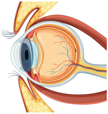
The cornea is the clear window at the front of your eye, over the coloured iris. The sclera is the white part around it. Anything sitting on the clear window deserves more attention than something on the white.
7 Causes of a White Spot on the Eye
1. Pinguecula
A pinguecula is a small, raised, white or yellowish growth on the conjunctiva, the clear membrane covering the white of the eye. It usually appears on the side nearest the nose.
It develops from long-term ultraviolet exposure and is made worse by dryness, wind and dust. It may contain deposits of protein, fat or calcium.
For most people it does not affect vision. If it becomes raised enough to disturb the tear film, it can cause dryness, redness and irritation. Lubricating drops usually settle this. Removal is uncommon.
2. Pterygium
A pterygium is a raised, wedge-shaped growth of the conjunctiva that extends onto the cornea. That crossing onto the clear window is what separates it from a pinguecula.
The same causes apply: UV exposure, dryness, wind and dust, which is why it is common in people who work outdoors. It typically causes redness, irritation and a gritty feeling.
A small pterygium may need nothing more than lubricants and sun protection. A larger one can change the curvature of the cornea and distort vision, and may be removed surgically. It can recur months or years after removal.
3. Corneal ulcer (infective keratitis)
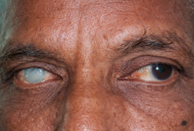
This is the cause that changes the urgency of everything else. A corneal ulcer is an open sore on the cornea, usually from infection, and it commonly appears as a white or greyish spot on the clear part of the eye.
It is rarely subtle. Expect some combination of:
- Eye pain, often significant
- Marked redness
- Watering
- Sensitivity to light
- Blurred vision
- A feeling that something is in the eye
- Discharge
Common routes in are contact lens misuse, an eye injury, and a scratch from vegetation. A corneal ulcer can scar the cornea permanently and threaten vision, and treatment depends entirely on identifying the organism responsible. This needs same-day examination, not a wait-and-see week.
4. Corneal scar or opacity
A corneal scar is what an ulcer, injury or old infection can leave behind: a permanent white or cloudy patch on the cornea.
Unlike an active ulcer, a scar is painless, stable and does not change from week to week. Whether it affects vision depends entirely on position: a scar away from the centre may cause no symptoms at all, while one directly over the pupil can blur vision significantly.
Scars do not clear on their own. Where vision is affected, options range from specialist contact lenses to corneal surgery, and depend on depth and location.
5. Conjunctival concretions
These are tiny, hard, white or yellowish specks that form in the lining of the eyelid. They are usually noticed only when the lid is pulled back, and they are more common with increasing age and long-standing lid inflammation.
Most cause nothing at all. Occasionally one works its way to the surface and produces a persistent scratchy, foreign-body sensation, and can then be removed easily in clinic.
6. Growths that need to be checked
Not every raised white lesion on the surface of the eye is a pinguecula. Some pre-cancerous growths of the ocular surface can look very similar, which is why an eye specialist examines them under a slit-lamp microscope and occasionally removes a growth so it can be examined under a laboratory microscope.
This is not a reason for alarm. It is a reason not to self-diagnose a lump as harmless because it resembles pictures you have seen online. Get a growth on the surface of the eye looked at once, particularly if it is enlarging, changing colour, developing its own blood vessels, or bleeding.
7. A white reflex in the pupil (leukocoria)
This one is different from the other six. Here the whiteness is not on the surface of the eye at all it is coming from deep inside, showing through the pupil.
In adults, the most common reason is an advanced cataract, where the lens has become dense and pale.
In children, a white pupillary reflex is a recognised warning sign that requires prompt ophthalmic examination. It is covered in full in the next section.
The Two Causes That Cannot Wait
A painful white spot on the cornea
If you have a white spot on the clear part of your eye together with pain, redness, watering, light sensitivity or blurred vision, treat it as urgent and get to an eye hospital the same day.
Corneal infection can progress quickly. The cornea has no blood supply of its own, which limits how well the body fights infection there, and scarring in the visual axis is permanent. Identifying whether the cause is bacterial, fungal or viral requires examination and often a laboratory sample, and the treatments are completely different.
Contact lens wearers should remove their lenses immediately, keep the lenses and the case, and bring both to the appointment.
A white pupil in a child
If a child’s pupil appears white, pale or shows no red reflex, arrange an eye examination promptly. Clinical guidance points to assessment by an eye specialist within about a week.
Two practical points that matter more than any description:
It is very often first noticed in a photograph. In a flash photo, healthy pupils both glow red. If one pupil looks red and the other looks white, pale or simply darker, that asymmetry is worth acting on. Many parents notice it in a family picture before anyone notices it in daylight.
A squint can appear alongside it. A child who develops a new inward or outward turn of one eye, with or without a white reflex, needs examination rather than reassurance that they will grow out of it.
The reason for urgency is that the causes of a white pupil in a child include serious conditions of the retina and lens, including retinoblastoma, the most common eye cancer of childhood. It is highly treatable when found early, and Indian data has shown that delay between a parent first noticing something and reaching specialist care is associated with worse outcomes.
Two things are worth saying alongside that. Most children referred for an abnormal red reflex turn out to have nothing serious; referral is still the right response. And do not attempt any tests or treatments first: the correct step is examination by an eye specialist, not a trial of drops.
Why Corneal Infection Deserves Extra Caution in India
Corneal injury from plant material is a recognised route into serious eye infection here: a whip from a tree branch, sugarcane leaf, crop debris, or dust thrown up during farm work.
These injuries matter because vegetative matter can introduce fungal infection, which behaves differently from bacterial infection: it develops more slowly, is harder to identify without laboratory testing, and requires antifungal rather than antibacterial treatment.
There is one specific danger. Steroid eye drops make fungal corneal infection significantly worse, and steroid drops are widely available. Somebody with a painful white corneal spot who is given, or reuses, a steroid drop can convert a treatable infection into a sight-threatening one.
If you work outdoors or in farming and you get anything in your eye, protective eyewear beforehand and a prompt examination afterwards are the two things that prevent most of these outcomes.
What Not to Do While You Wait for an Appointment
- Do not use leftover eye drops from a previous problem or a family member’s prescription, particularly steroid drops. On an undiagnosed white corneal spot this can cause real harm.
- Do not buy an over-the-counter “eye infection” drop and hope. Bacterial, fungal and viral infections need different medicines, and the wrong one wastes days.
- Do not rub the eye.
- Do not patch the eye unless an eye specialist has told you to.
- Remove contact lenses and do not put them back until you have been examined.
- Do not use home remedies such as rose water, breast milk, honey, kajal or herbal preparations on an eye with a white spot, pain or discharge.
- Do not wait for it to clear on its own if there is pain, redness or any change in vision.
How an Eye Specialist Identifies the Cause
- History – how long the spot has been there, any injury, contact lens use, work exposure, and whether vision has changed.
- Vision check – measured for each eye separately.
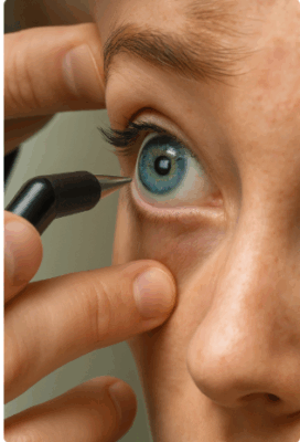
- Slit-lamp examination – a microscope that shows the layers of the cornea and conjunctiva in detail, and is the single most informative step.
- Fluorescein staining – a harmless orange dye that glows under blue light and reveals breaks in the corneal surface, distinguishing an active ulcer from an old scar.
- Corneal scraping – where infection is suspected a small sample is sent for laboratory testing to identify the organism.
- Dilated retinal examination – if the whiteness is coming from inside the pupil rather than the surface.
- Imaging or ultrasound – in selected cases, particularly when the view inside the eye is obscured.
Most patients need only the first three steps.
When to See a Doctor
Go the same day if you have:
- A white spot on the clear part of your eye with pain, redness or discharge
- Any sudden drop in vision
- Marked sensitivity to light
- A white spot appearing after an eye injury, especially from plant material
- A white spot while wearing contact lenses
- A spot that is enlarging over days
Arrange an appointment within days if you notice:
- A white or pale reflex in a child’s pupil urgently, in a child
- A new growth on the surface of the eye that is changing
- Persistent irritation from a longstanding spot
- A white pupil in an adult with gradually worsening vision
A routine appointment is reasonable for:
- A small, stable white or yellow bump on the white of the eye with no pain
- A longstanding corneal scar that has not changed and does not affect vision
Also relevant: inflammation inside the eye can produce a white or yellowish level at the bottom of the coloured part of the eye. That combination of whiteness inside the eye with pain and light sensitivity is another same-day presentation.
Conclusion
If you take one thing from this article, make it the location check. A white spot on the white of the eye is usually a UV-related growth that needs sun protection and lubricating drops. A white spot on the clear cornea with pain needs an eye hospital today. A white reflex inside a child’s pupil needs a specialist this week.
The mistake that causes the most harm is not delay in itself it is reaching for a drop first. Steroid drops in particular can turn a treatable corneal infection into a permanent scar, and no drop is a substitute for finding out what the spot actually is.
If you have noticed something on your eye and are not sure which category it falls into, that uncertainty is itself a reason to book a comprehensive eye examination. A slit-lamp examination takes minutes and settles the question.