


Most people have floaters. Those drifting specks, threads and cobwebs that swim across your vision against a bright sky or a white wall are, for the vast majority, a harmless part of the eye ageing.
The situation changes when they are new. A sudden shower of floaters, or flashes of light appearing at the edge of your vision, can be the only warning that the retina has torn, and a torn retina that goes untreated can detach and cost you sight in that eye. This guide explains how to tell the difference and what to do.
Key Takeaway: Are Flashes and Floaters Serious?
Long-standing, stable floaters are almost always harmless. New flashes and floaters are not an emergency in most people either, but roughly 5 to 10 percent of those who develop them acutely are found to have a retinal tear, which is why the symptoms need a dilated eye examination within days. Go to an eye casualty or emergency department immediately if you see a curtain or shadow spreading across your vision, a sudden dense shower of floaters, or any loss of vision.
What Are Floaters and Flashes?
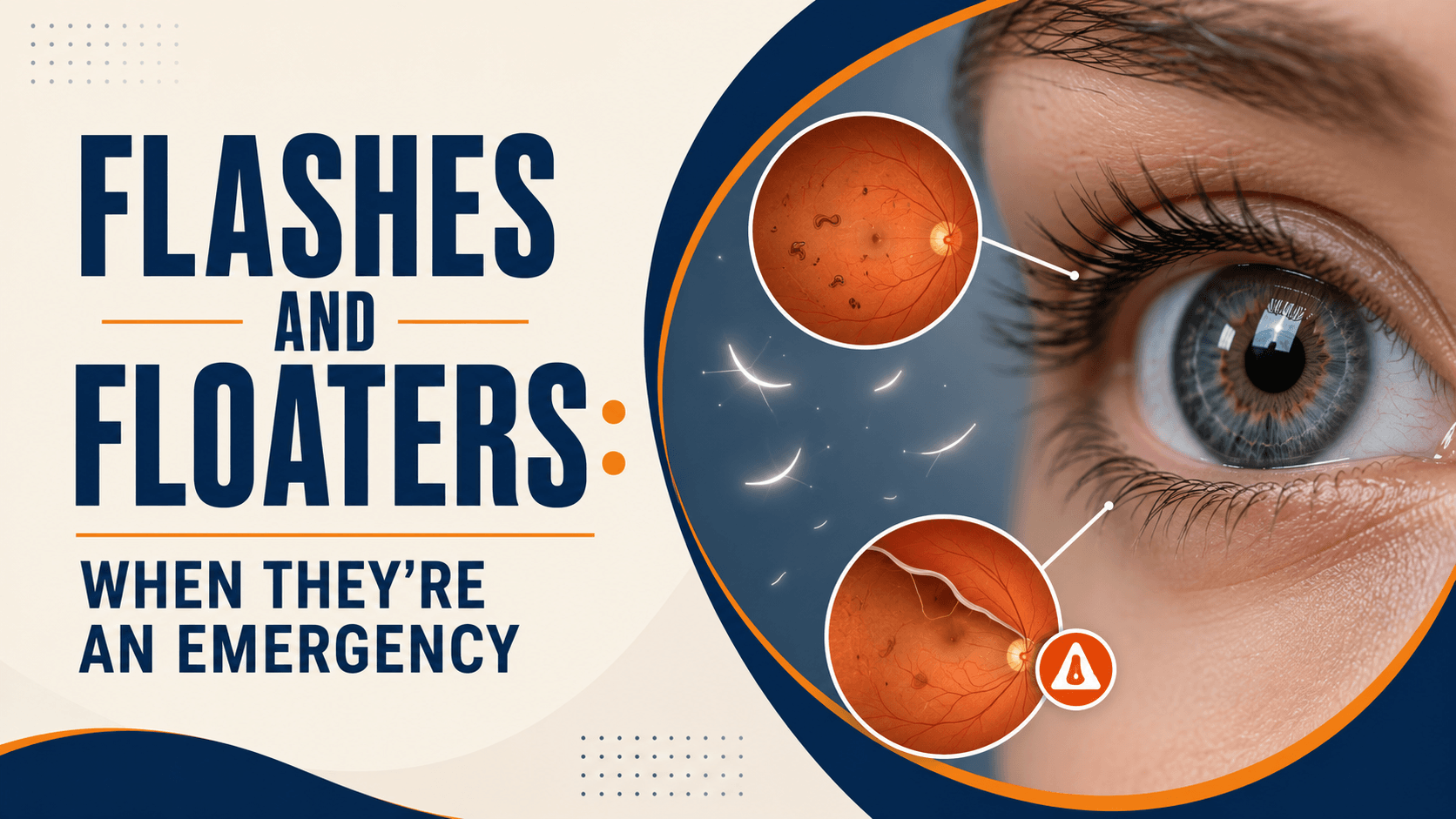
The inside of your eye is filled with a clear gel called the vitreous. With age, this gel liquefies and small clumps of collagen form within it. These clumps cast shadows on the retina, and those shadows are what you see as floaters. They drift as your eye moves, and they are usually most obvious against plain, bright backgrounds.
Flashes, known medically as photopsia, come from a different mechanism. As the vitreous gel shrinks, it can tug on the retina. The retina cannot feel pulling; it only understands light, so it interprets that mechanical tug as a flash, often described as a brief arc or streak of light at the edge of vision, more noticeable in the dark.
That distinction matters. Floaters are usually about debris. Flashes are about traction something pulling on the retina, which is why new flashes deserve more attention than new floaters alone.
The Most Common Cause: Posterior Vitreous Detachment
Most new flashes and floaters are caused by posterior vitreous detachment (PVD), in which the vitreous gel separates from the retina at the back of the eye. It is a normal age-related event, most common after 50, and by itself it is not a disease.
A typical PVD announces itself with a sudden increase in floaters often one prominent large floater, sometimes described as a ring or a fly along with flashes for a few weeks. Over the following months, the symptoms usually settle, and the brain gradually learns to ignore the remaining floaters.
The complication is what matters. As the gel pulls away, it can tear the retina where the two are firmly stuck together. That is why a normal, common, age-related process still needs checking: the PVD is harmless, but the tear it can cause is not.
Once you have had a PVD in one eye, the other eye usually goes through the same process within six months to two years. Knowing this in advance makes the second episode far less frightening.
When Flashes and Floaters Are an Emergency
Seek emergency eye care the same day if you experience:
- A curtain, shadow or veil spreading across any part of your vision
- A sudden dense shower of floaters, or floaters like smoke, soot or a swarm
- Loss of vision in any part of your visual field
- Flashes that are increasing in frequency or intensity rather than settling
- New floaters or flashes after an eye injury or head trauma
- New symptoms following recent eye surgery
- Floaters or flashes with eye pain or redness
The curtain or shadow is the most important of these. It usually means the retina has already begun to detach, and the time to treatment strongly influences how much vision is saved, particularly if the central macula has not yet lifted.
The sudden dense shower matters for a different reason: it often indicates bleeding into the vitreous, and when a PVD is accompanied by vitreous haemorrhage the likelihood of an underlying retinal tear is substantially higher than in an uncomplicated PVD.
Why New Symptoms Need Checking Within Days
Even without the emergency features above, new flashes and floaters warrant a dilated retinal examination within a few days.
The reason is straightforward. Across large studies of people presenting with acute symptomatic posterior vitreous detachment, retinal tears are found in roughly 5 to 10 percent, with a smaller proportion already showing a detachment. In other words, most people are fine, but around one in ten to one in twenty are not, and there is no way to tell from symptoms alone which group you are in.
The second reason is the treatment window. A symptomatic retinal tear that is still being pulled on carries a high risk of progressing to a full retinal detachment if left alone, reported at around half of such cases. Sealing a tear is a quick outpatient laser procedure. Repairing a detachment is major surgery with a far less certain visual outcome. The gap between those two situations is often only a matter of days.
That is the entire argument for going promptly: not because the odds are bad, but because the downside of being in the unlucky minority is severe and the fix while it is still a tear is simple.
What Is Usually Not an Emergency
Equally important, so you are not alarmed unnecessarily:
- Floaters you have had for years that have not changed
- A few small floaters noticed occasionally against bright backgrounds
- Floaters that are gradually becoming less noticeable
- Brief coloured or zigzag patterns affecting both eyes, lasting 20 to 60 minutes and often followed by a headache, which are more typical of migraine visual aura than a retinal problem
- Seeing tiny moving dots in bright blue sky, which is a normal optical phenomenon
If your floaters have been stable for a long time and nothing has changed, they should still be mentioned at your routine eye check, but they do not require an urgent appointment.
Other Causes of Flashes and Floaters
|
Cause |
Typical features |
Urgency |
|
Posterior vitreous detachment |
New floaters plus flashes, usually after 50 |
Examination within days |
|
Retinal tear |
Same as PVD, sometimes with a shower of floaters |
Urgent same day if possible |
|
Retinal detachment |
Curtain or shadow, loss of visual field |
Emergency |
|
Vitreous haemorrhage |
Sudden dense floaters, smoky or red haze, reduced vision |
Emergency |
|
Floaters with eye pain, redness, light sensitivity |
Prompt assessment |
|
|
New floaters in someone with diabetes, from bleeding |
Urgent |
|
|
Migraine visual aura |
Zigzag or shimmering shapes, both eyes, 20–60 minutes |
Not urgent once diagnosed |
Who Is at Higher Risk?
Some people are considerably more likely to develop a retinal tear when a PVD occurs:
- Short-sightedness, especially high myopia; the eye is longer and the retina thinner and more stretched. Given how rapidly myopia is increasing in India, this puts a growing number of younger adults at risk, most of whom have never been told.
- Previous cataract surgery or other intraocular surgery
- Previous retinal tear or detachment, in either eye
- A family history of retinal detachment
- Eye injury, including sports and workplace trauma
- Lattice degeneration, a thinning of the peripheral retina often found incidentally
If any of these apply to you, treat new flashes or floaters with more urgency, not less, and make sure the person examining you knows your history.
What Happens at Your Eye Examination
The examination is painless and usually takes under an hour, most of which is waiting for the drops to work.
- Vision and pressure checks, and a history of exactly when and how symptoms started.
- Dilating drops, which take 20 to 30 minutes to widen the pupil. Vision will be blurred and light-sensitive for several hours afterwards.
- Slit-lamp examination of the front of the eye and the vitreous, looking for pigment cells or blood, both of which raise suspicion of a tear.
- Indirect ophthalmoscopy with scleral indentation, where the doctor examines the far periphery of the retina. This part is essential; tears usually occur at the edge, beyond what a standard examination reveals.
- Both eyes are examined, not only the symptomatic one.
- Scans where needed, including OCT, or ultrasound if bleeding blocks the view of the retina.
Arrange for someone to accompany you and do not plan to drive, as your vision will be blurred by the drops for some hours.
Treatment for Retinal Tears and Detachment
If a tear is found, sealing it is usually quick and done the same day.
- Laser retinopexy – a retinal laser creates small burns around the tear that scar down and weld the retina to the underlying tissue, preventing fluid from getting underneath. It is done in the clinic under local anaesthetic and takes minutes.
- Cryopexy – a freezing probe applied to the outside of the eye achieves the same seal, and is used when a laser cannot reach or view the tear.
If the retina has already detached, treatment becomes surgical:
- Pneumatic retinopexy – a gas bubble is injected to push the retina back, combined with laser or freezing, in selected cases
- Scleral buckle – a silicone band supports the eye wall from outside
- Vitrectomy – the vitreous gel is removed and the retina repositioned, often with a gas or oil bubble
Outcomes depend heavily on whether the central macula was still attached when surgery took place, which is the clearest reason not to wait.
Can Ordinary Floaters Be Treated?
For most people, the honest answer is that no treatment is needed or advisable. Floaters typically become less noticeable over months, partly because they settle and partly because the brain learns to filter them out.
Where floaters are genuinely disabling, two options exist, and both carry real trade-offs:
- Vitrectomy removes the vitreous gel and the floaters with it. It is effective, but it is intraocular surgery carrying risks including cataract formation, retinal detachment and infection. It is generally reserved for severe, persistent cases after careful discussion.
- Laser vitreolysis breaks up floaters with a laser. Evidence for benefit is limited, results vary, it is unsuitable for many floater types, and it is not risk-free.
Be cautious of any product or clinic promising to dissolve floaters with drops, supplements or exercises. No such treatment is supported by good evidence.
Why One Normal Examination Is Not the End of It
This is the part patients are least often told, and it matters.
A clear examination today does not guarantee a clear examination in three weeks. Because the vitreous is still separating, a proportion of people whose first check is normal develop a retinal tear in the following weeks. For this reason, a follow-up dilated examination is commonly arranged a few weeks later after an acute PVD.
More importantly, you should return immediately – without waiting for that appointment – if your flashes increase, your floaters suddenly multiply, or any shadow appears. Those symptoms override any reassurance from a previous visit.
And because the fellow eye usually undergoes the same process within six months to two years, the same rules apply when it starts.
What to Do Right Now
- Check for emergency signs – a curtain or shadow, a sudden dense shower of floaters, or any vision loss. If present, go to emergency eye care now.
- Cover each eye in turn and note which eye the symptoms are in, and roughly which part of your vision. This genuinely helps your doctor.
- Note when it started and whether it is getting worse.
- Book a dilated eye examination within a few days, even if you feel fine.
- Tell them your history – short-sightedness, previous eye surgery, injury, or family history of detachment.
- Arrange transport, since you will not be able to drive after dilation.
- Do not wait to see whether it settles. With retinal tears, waiting is the only decision that cannot be undone.
Conclusion
The reassuring reality is that most floaters are harmless and most new flashes and floaters are an ordinary vitreous detachment that settles on its own. The reason this symptom still deserves respect is the minority, roughly one in ten to one in twenty with a retinal tear, who cannot be identified without dilating the pupil and examining the edge of the retina.
Remember the two rules. New symptoms need a dilated examination within days. And a curtain, a shadow, a sudden shower of floaters or any loss of vision means going now, not tomorrow.