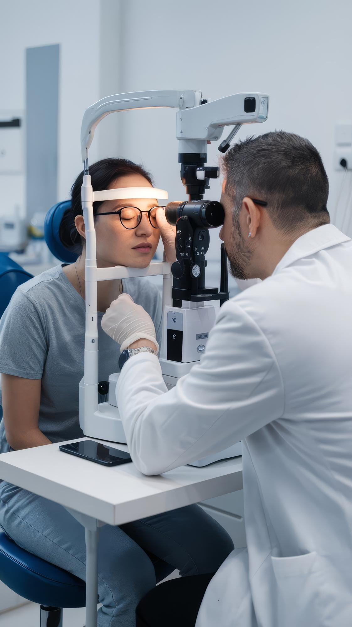
The Hirschberg test offers one of the quickest ways to check whether both eyes point in the same direction. Eye doctors also call it the corneal light reflex test. During the test, the clinician shines a small light into the patient’s eyes and observes the tiny reflection on the cornea. This reflection is known as the first Purkinje image.
In everyday clinical practice, the Hirschberg eye test helps detect strabismus, or misalignment of the eyes. Pediatric ophthalmologists rely on it frequently because young children rarely cooperate with detailed eye movement testing. The Hirschberg test helps confirm whether this appearance reflects real misalignment or simply facial anatomy.
Understanding the Hirschberg Test
Definition and Purpose of the Hirschberg Test
The Hirschberg test serves as a screening tool for ocular alignment. The doctor directs a focused light toward both eyes while the patient looks straight at the light source. The clinician also calls this the corneal light reflex, as it assesses the cornea’s reflection of light.
Why and When the Hirschberg Test Is Used
In clinical practice, the Hirschberg eye test becomes extremely useful when a patient cannot cooperate with complex eye exams. Eye doctors commonly use the test in situations such as:
- Infants and toddlers who cannot follow instructions
- Patients with developmental delays
- Emergency examinations when quick screening is required
Anatomy and Optics Behind the Corneal Light Reflex
The Corneal Reflex and Purkinje Images
When light reaches the eye, it reflects off several surfaces within the eye. These reflections are known as Purkinje images. The Hirschberg test focuses on this image, which forms on the tear-film-covered cornea.
The cornea acts like a mirror, producing a small, bright reflection. During the Hirschberg test procedure, doctors examine this reflection carefully. If both eyes are properly aligned, the reflections appear in matching positions relative to each pupil.
Normal Alignment and Angle Kappa
In a normally aligned eye, the corneal reflex does not sit exactly at the centre of the pupil. Instead, it lies slightly toward the nose. This offset occurs because the visual axis differs slightly from the eye’s anatomical centre.
Doctors call this offset angle kappa. Angle kappa can influence how the eyes appear during the test:
- Positive angle kappa may create the appearance of exotropia
- Negative angle kappa may mimic esotropia
- A normal small nasal reflex usually indicates healthy alignment
How the Hirschberg Test Works
Step-By-Step Procedure
The Hirschberg test procedure follows a straightforward sequence.
Set up
- The examiner holds a penlight or transilluminator about 50-60 cm away from the patient.
- The room lighting remains slightly dim to improve visibility of the reflection.
Patient fixation
- The patient focuses directly on the light source.
- This step ensures both eyes attempt to fixate on the same target.
Observation
- The clinician observes the corneal reflex in both eyes.
- Symmetrical reflections indicate normal alignment.
- A shifted reflex suggests ocular deviation.
The entire process usually takes less than a minute.
Interpreting Results: Normal vs Misaligned
Doctors interpret the position of the corneal reflex to determine the type of misalignment.
- Orthotropia or normal alignment: Symmetric central reflex
- Exotropia or outward deviation: Reflex shifted nasally
- Esotropia or inward deviation: Reflex shifted temporally
- Hypotropia: Reflex shifted upward
- Hypertropia: Reflex shifted downward
Estimating Misalignment Using Millimetres and Prisms
The distance between the reflex and the pupil centre provides an estimate of the deviation angle. Clinicians use common approximations:
- 1 mm displacement ≈ 7 degrees or 15 prism diopters
- Reflex at pupillary margin (≈2 mm) ≈ 15 degrees or 30 prism diopters
- Reflex at mid-iris (≈4 mm) ≈ 30 degrees or 60 prism diopters
- Reflex at limbus ≈ 45 degrees or about 90 prism diopters
Hirschberg vs Krimsky Test
The Hirschberg and Krimsky tests share similar principles but differ in precision. Key differences include:
|
Feature |
Hirschberg Test |
Krimsky Test |
|
Method |
Visual estimation of deviation |
Uses prisms placed in front of the eye |
|
Precision |
Quick screening method |
Measures deviation more accurately |
Applications and Clinical Significance
Using the Test to Screen for Strabismus and Amblyopia
Eye specialists use the Hirschberg test widely during routine examinations. Paediatricians often perform it during early childhood checkups. The test helps screen for conditions such as:
- Strabismus
- Amblyopia (lazy eye)
- Cranial nerve abnormalities
- Neurological disorders affecting eye movement
How the Test Guides Management
The Hirschberg test often provides the first indication of misalignment. When doctors detect asymmetry, they conduct additional examinations. Further tests may include:
- Cover-uncover test
- Alternate cover test
- Prism cover measurements
- Comprehensive ocular motility evaluation
Early diagnosis allows doctors to initiate treatments such as glasses, rule out complications like amblyopia, and, occasionally, perform patching therapy or, when alignment problems become severe, eye surgery.
Hirschberg Test for Infants and Pseudostrabismus
Many parents worry when their baby appears cross-eyed. In reality, infants often show pseudostrabismus, which results from facial features such as:
- Flat nasal bridge
- Prominent epicanthal folds
- Wide nasal bridge
In these cases, the Hirschberg test becomes extremely helpful. If the corneal reflex appears centred in both eyes, alignment remains normal despite the appearance.
Conclusion: The Value of the Hirschberg Test
The Hirschberg test remains one of the simplest yet most valuable screening tools in ophthalmology. The test proves particularly useful for infants, young children, and patients who cannot cooperate with detailed eye movement exams. Although it does not replace more precise diagnostic tests, it plays a critical role in the early detection of strabismus and related conditions.