Key Takeaways
- Ashish, 35, a professor at the time, had vision problems with dark spots and flashing lights that affected his ability to work.
- He was found to have a retinal detachment and required a procedure called scleral buckle surgery to fix it.
- Scleral buckle surgery is a procedure that uses a sponge or buckle is placed against the eye to support the retina and may also involve other aspects in an effort to prevent tearing of the retina.
- Scleral buckles include three types: radial, encircling circumferential, and segmental circumferential, and are designated for differing types of retinal problems.
- After the procedure, Ashish had two to four weeks of recovery and was provided with aftercare.
As a renowned eye hospital, we have to skillfully address hundreds of patients who require comprehensive eye treatments. A couple of weeks ago, we met Ashish, a young 35-year-old professor who has been teaching sociology in a prestigious college at the University of Delhi. He was accompanied by his wife, who was constantly fidgeting with her purse, indicating nervousness.
When asked about the problem, we learned that Ashish had been witnessing small dark spots and sudden flashes of light in his left eye. Slowly, as the frequency of these symptoms increased, he faced difficulty in reading and writing, which forced him to take regular leaves from college.
When we asked him to describe his symptoms more elaborately, he mentioned blurred vision, reduced peripheral vision, and recalled temporary vision loss in this right eye. We noted down the symptoms, asked him to walk us through his medical history and told him to visit us the next day.
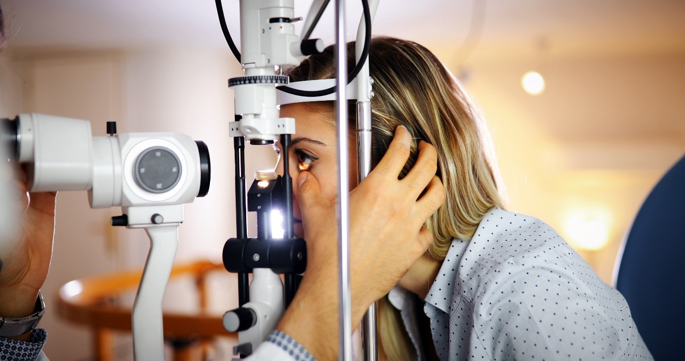
In the next session, we sat him through a detailed diagnostic procedure by conducting ultrasound imaging and retinal examination. After a day or two, when the results pointed towards retinal detachment, we sat Ashish and his wife down and explained that Ashish had to undergo a scleral buckle surgery. To give them more clarity, we calmly explained to them the fundamental definition of retinal detachment and scleral buckle surgery in simple layperson terms.
Retinal detachment is referred to as an eye problem that occurs when the retina is displaced or pulled away from its normal position at the back of the patients’ eye. Rhegmatogenous, tractional, and exudative are three types of retinal detachment that can be caused by ageing, hereditary, serious eye injury, severe nearsightedness, and more.
An Insight into Scleral Buckle Surgery: Steps and Procedure
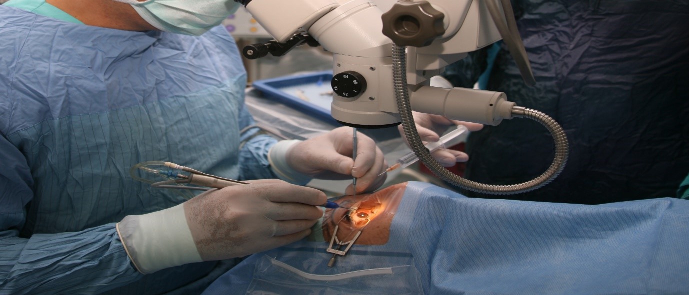
Scleral buckle surgery is a minute procedure carried out by skilled surgeons and doctors in the ophthalmological field. Before the surgery, we asked Ashish to lay back, relax, and, step by step, walk him through the process of the surgery so that he gains a sense of confidence and comfort.
In the first couple of minutes before the scleral buckle surgery, we gave him two options. The first one was sedating him with general anaesthesia, which would put him asleep through the procedure and the second, he could choose to stay awake. In this case, the doctor will give you eye drops or an injection to numb your eye. Now, let us have a glimpse into how scleral buckle surgery is carried out:
- First and foremost, the patient is given eye drops to widen the pupil. This allows the doctor or the surgeon to see the back of the patient’s eye.
- In the second step, with the use of high-quality medical tools and technology, an incision is made in the outer layer of the eye, also called the sclera.
- Now, a sponge or a buckle is surgically sewn around the outer layer of the eye to ensure that it doesn’t move from the allotted spot. The idea of buckling is designed to optimally support the retina by pushing the outer layer of the eye towards the middle, this can reattach the retina and close all retina tears if there are any.
- To be doubly sure that a tear does not reopen, the surgeon might perform one of the following procedures:
- Cryopexy: In this process, the surgeon freezes the sclera, which can often lead to the development of scar tissue.
- Laser photocoagulation: In order to burn the area around a retinal detachment or tear, the doctor might deploy a laser beam. This results in scar tissue which stops fluid leakage and helps seal a break.
- Once the primary steps of the scleral buckle surgery are over, the doctor drains any residual fluid behind the retina and gives the patient anti antibiotic eye drops to prevent any kind of infection.
Scleral Buckle Types and Precautions: A Brief Overview
Depending on the patient’s situation, this surgery can be divided into three scleral buckle types. In order to receive the best scleral buckle surgery, it is best to visit a renowned and trustworthy hospital that has competent ophthalmologists to perform the right diagnosis. One by one, let’s delve into the above-mentioned scleral buckle types:
- Radial Buckle: This is only used in a single retinal break with a significant flap tear. Additionally, this type of buckle is also used in cases where the tear exhibits rolled edges or have irregularities.
- Encircling Circumferential Buckle: The surgeon decides to employ this type of buckle when the retinal breaks in more than three quadrants or where there is a chance of unidentifiable retinal breaks.
- Segmental circumferential buckle: In this type, the key premise is that only the tears causing the retinal detachment needs to be optimally supported by the buckle.
Once Ashish underwent the scleral buckle surgery, the recovery time took around two to four weeks. Since he was eager to go back to his students, we gave him a list of precautions and aftercare instructions like:
- Try to wear sunglasses during the day.
- Don’t drive unless allowed by the doctor.
- Keep in mind to take post-surgery medicines regularly and on time.
- Wear swimwear glasses while taking a shower to prevent water or soup from entering the eye.
- Avoid rapid eye movements and stay in touch with your doctor in case you sense any discomfort.
Dr Agarwals Eye Hospital: Offering Best-in-Class Eye Solutions Since 1957
At Dr Agarwals Eye Hospital, we have equipped our medical faculty with top-notch technology and infrastructure. In addition to scleral buckle surgery, we also offer treatments like glued IOL, oculoplasty, refractive surgery, photorefractive, and more.
With 100+ hospitals across 11 countries, we are known for seamlessly combining experience with exceptional knowledge to provide a wide variety of eye solutions. Book an appointment today to receive an unmatched hospital experience with personalized care!