



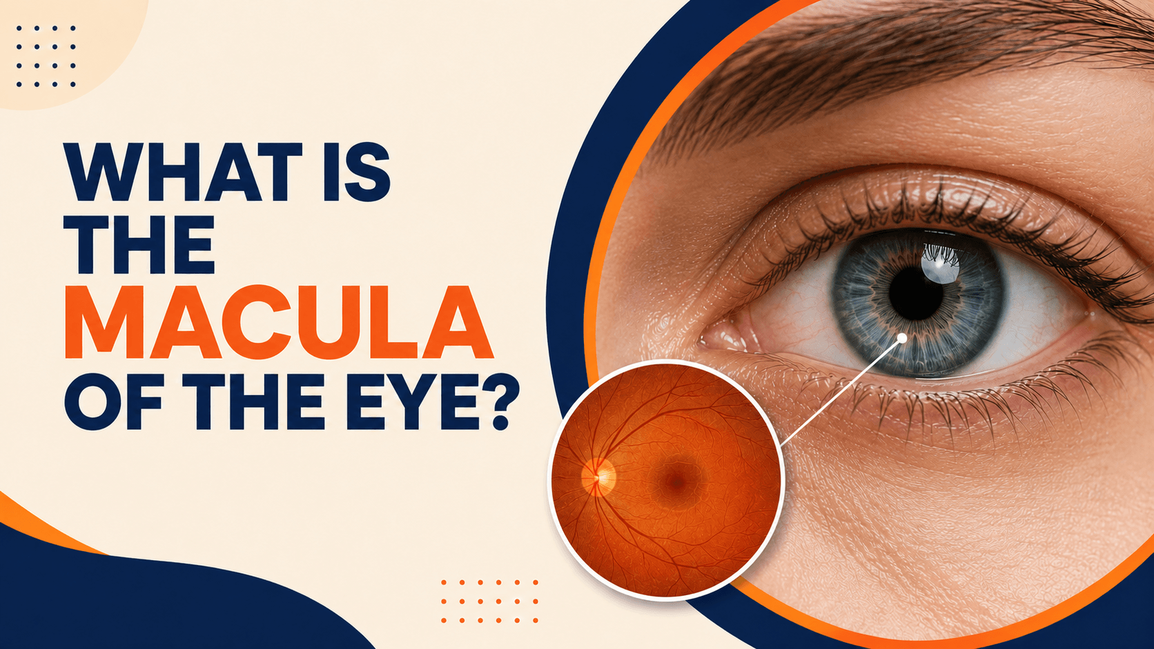
The macula of the eye is the small, central, yellowish part of the retina responsible for your sharp, detailed vision. Although it makes up only a tiny portion of the retina, the macula does the heavy lifting for the tasks that matter most every day reading, recognising faces, seeing fine detail, and perceiving colour. When the macula is healthy, your central vision is crisp and clear; when it is damaged, those everyday tasks can become difficult. This guide explains the macula’s definition, structure, function, and the common disorders that can affect it.
Understanding the Macula: Location & Structure
To understand the macula definition fully, it helps to know where it sits. The macula is located near the centre of the retina, at the back of the eye, roughly in line with the centre of the pupil. It is sometimes called the macula lutea, meaning “yellow spot,” because of the yellow pigments (lutein and zeaxanthin) concentrated there.
The macula is small only about 5 to 6 mm across yet it is densely packed with photoreceptor cells, the light-sensing cells of the retina. The macula is especially rich in cone cells (which handle detailed, colour, and daytime vision), with rod cells (for dim-light vision) increasing toward its edges. At the very centre of the macula is a tiny pit called the fovea, and at the heart of the fovea lies the foveola the point of your sharpest possible vision, made up almost entirely of cones.
What Does the Macula Do? – Key Functions
The key function of the macula is to give you central vision the focused, detailed sight you use when you look directly at something. The photoreceptors in the macula capture incoming light and convert it into electrical signals, which travel along the optic nerve to the brain, where they are interpreted as images.
Because of its dense cone population, the macula is responsible for:
- Reading small text and seeing fine detail
- Recognising faces and expressions
- Discriminating colours accurately
- Tasks needing precise central vision, such as driving and threading a needle
The rest of the retina handles your peripheral (side) vision, which is why macular problems typically affect central vision while side vision often stays intact.
Signs and Symptoms of Macular Problems
Macula symptoms usually involve the centre of your sight. Recognising them early is important, because prompt treatment can preserve vision. Common warning signs include:
- Blurred or dim central vision
- Distorted vision, where straight lines appear wavy or bent
- A dark, blurry, or blank spot in the centre of your sight
- Difficulty recognising faces
- Needing brighter light to read
- Colours appearing less vivid
- Sensitivity to light (photophobia)
- Sudden vision loss, which is a medical emergency
If you notice any of these changes especially blurred central vision or sudden distortion see an eye specialist promptly rather than waiting for symptoms to worsen.
Common Macular Disorders & Diseases
Several macular disorders specifically affect this central area of the retina. The most common macular diseases include age-related macular degeneration, macular pucker (epiretinal membrane), macular hole, and cystoid macular edema. Other retinal conditions such as retinal detachment, retinal vein occlusion, and retinitis pigmentosa can also affect macular health.
Age-Related Macular Degeneration (AMD)
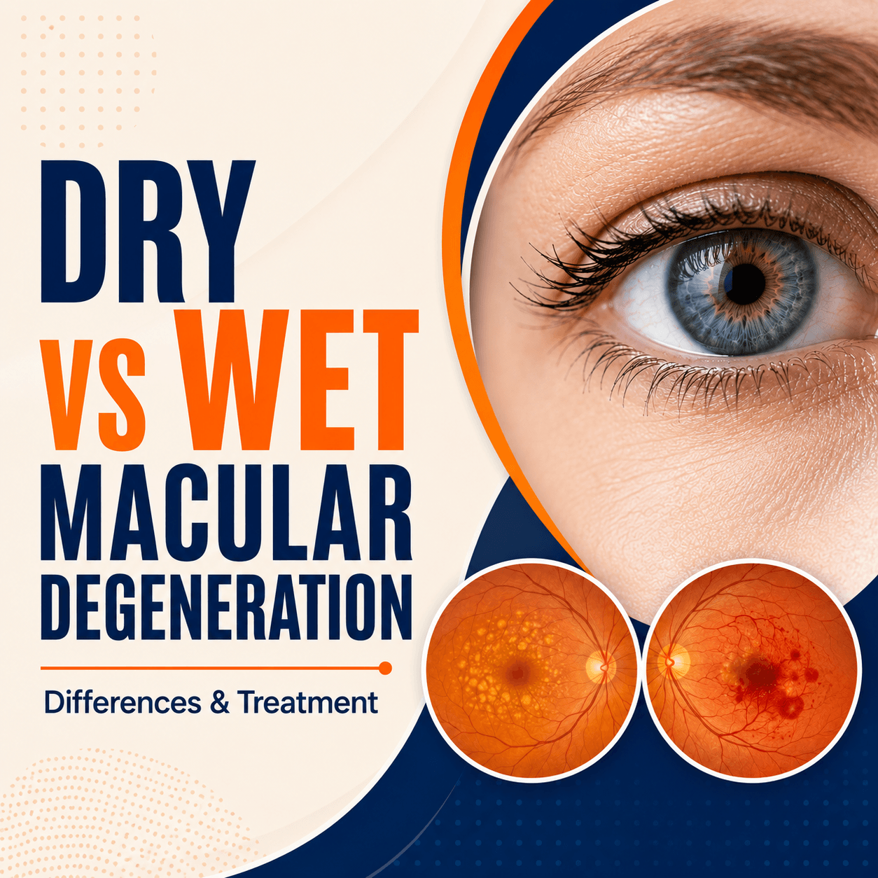
Macular degeneration is the most common macular disease and a leading cause of central vision loss in older adults. There are two forms. Dry AMD is the more common type, marked by tiny yellow deposits called drusen and gradual thinning of the macula. Wet AMD is less common but more aggressive, caused by abnormal blood vessel growth beneath the retina (choroidal neovascularisation) that leaks fluid or blood. Key risk factors for age-related macular degeneration include increasing age, smoking, genetics and family history, high blood pressure, and obesity.
Macular Hole
A macular hole is a small break in the centre of the macula. It often develops when the vitreous gel inside the eye pulls on the macula (vitreous traction) as part of ageing, and it can also follow an eye injury. A macular hole typically causes blurred or distorted central vision and progresses through stages, from an impending hole to a full-thickness hole. Surgery a vitrectomy is used to close the hole and improve vision.
Macular Pucker (Epiretinal Membrane)
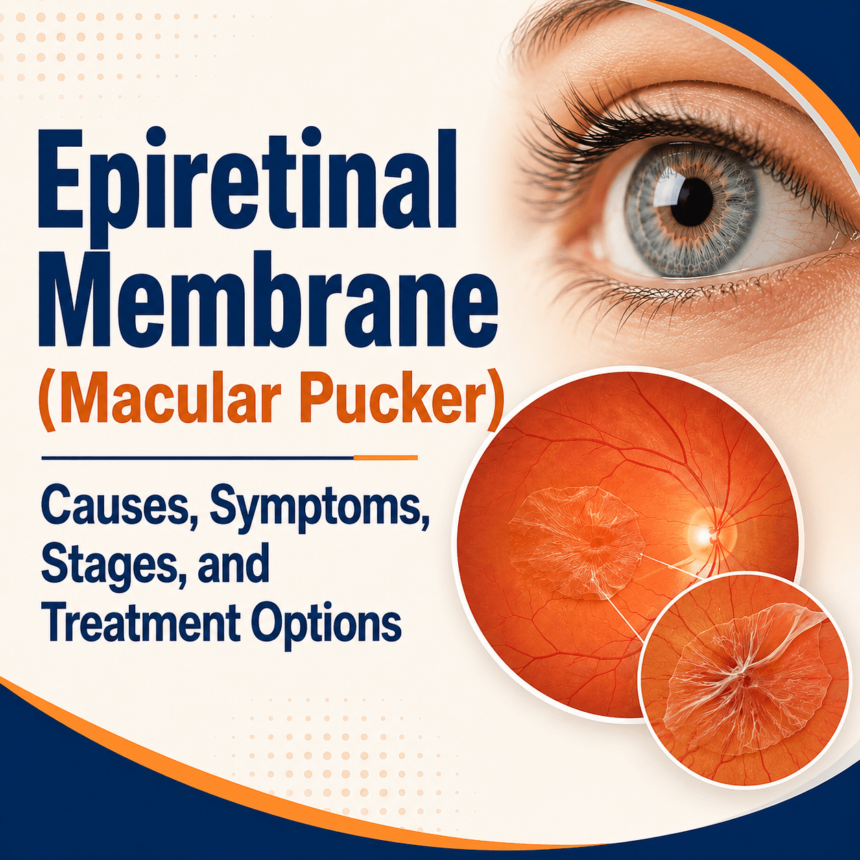
A macular pucker, also called an epiretinal membrane, is a thin layer of scar-like tissue that forms on the surface of the macula. As it contracts, it wrinkles the retina, leading to blurred and distorted central vision. Mild cases are simply monitored, while a vitrectomy may be recommended if vision is significantly affected.
Cystoid Macular Edema & Other Conditions
Cystoid macular edema is the build-up of fluid in cyst-like spaces within the macula, causing swelling and blurred vision. It can occur after cataract surgery, or as a result of diabetic retinopathy, retinal vein occlusion, or inflammation inside the eye. A related and common condition is diabetic macular edema, a major cause of vision loss in people with diabetes. Inherited conditions such as macular dystrophies (for example, Best disease) and other rarer disorders can also affect the macula.
Diagnosis: How Macular Conditions Are Detected
Diagnosing macular conditions is painless and relies on a combination of tests:
- Dilated retinal examination using an ophthalmoscope to view the macula directly
- Optical Coherence Tomography (OCT) — a quick, non-invasive scan that gives a detailed cross-section of the macula and is central to diagnosing most macular diseases
- Fluorescein angiography, which uses a dye to highlight abnormal or leaking blood vessels, especially in wet AMD
- Amsler grid, a simple self-test grid you can use to detect distortion in your central vision
- Visual acuity tests to measure how well you see
Early screening matters. People over 50, those with diabetes, and anyone with a family history of macular disease should have regular eye exams so problems are caught early.
Treatment Options for Macular Disorders
Macula treatment depends entirely on the specific condition and its severity. The aim is usually to preserve vision and slow progression. Options range from lifestyle measures and nutritional supplements (such as AREDS2 vitamins) to anti-VEGF injections, laser therapy, photodynamic therapy, vitrectomy surgery, and corticosteroid implants. Because every macular disease is different, treatment plans are tailored to the individual, and regular follow-up is essential.
Treating Age Related Macular Degeneration
Management differs by type. For dry AMD, there is no cure, but progression can often be slowed with a diet rich in lutein and zeaxanthin, stopping smoking, and AREDS2 supplements for certain stages of the disease. For wet AMD, the main treatment is anti-VEGF injections, which reduce abnormal blood vessel leakage; photodynamic therapy is sometimes used as well. Emerging approaches such as gene therapy and stem cell research are being studied and may offer additional options in the future.
Treating Macular Hole & Pucker
The standard treatment for a macular hole, and for a visually significant macular pucker, is a vitrectomy with membrane peeling, often combined with a gas bubble (gas tamponade) to help the macula heal. Many patients regain useful central vision, though recovery is gradual over weeks to months. A mild macular pucker may not require surgery at all monitoring and vision aids may be enough.
Treating Macular Edema & Other Conditions
Macular edema is treated by reducing the swelling and addressing its cause. Options include anti-VEGF therapy, steroid injections or implants, anti-inflammatory eye drops, and laser treatment, along with controlling underlying conditions such as diabetes or vein occlusions. For diabetic macular edema, injections are a common and effective approach. Timely intervention gives the best chance of preserving vision.
Living with Macular Disease: Prevention & Lifestyle
While not every macular condition is preventable, healthy habits help protect your macula and overall eye health:
- Wear UV-blocking sunglasses outdoors
- Eat a diet rich in leafy greens, fish, and antioxidants
- Control your blood pressure and blood sugar
- Stop smoking
- Maintain a healthy weight
- Have regular comprehensive eye exams
For those already living with vision loss, assistive devices such as magnifiers and large-print materials, along with vision rehabilitation services, can make a meaningful difference to daily life.
Conclusion: Protecting Your Central Vision
The macula may be tiny, but it is essential to almost everything you do with your eyes reading, recognising loved ones, and seeing the world in detail and colour. Protecting your central vision comes down to two things: early detection and healthy habits. If you notice any change in your central vision, such as blurring or distortion, seek timely care from an eye specialist. Combined with regular eye exams and a macula-friendly lifestyle, early action is the best way to keep your central vision sharp for years to come.






Very good analysis,I am under treatment and it’s better