


The eye is roughly the size of a table tennis ball, yet it performs a task no camera has fully matched: gathering light, focusing it with precision, converting it into electrical signals and sending them to the brain continuously and without you thinking about it.
This guide walks through a human eye diagram part by part, explains what each structure does, and shows which common eye conditions affect which part.
Key Takeaway: What Are the Main Parts of the Eye?
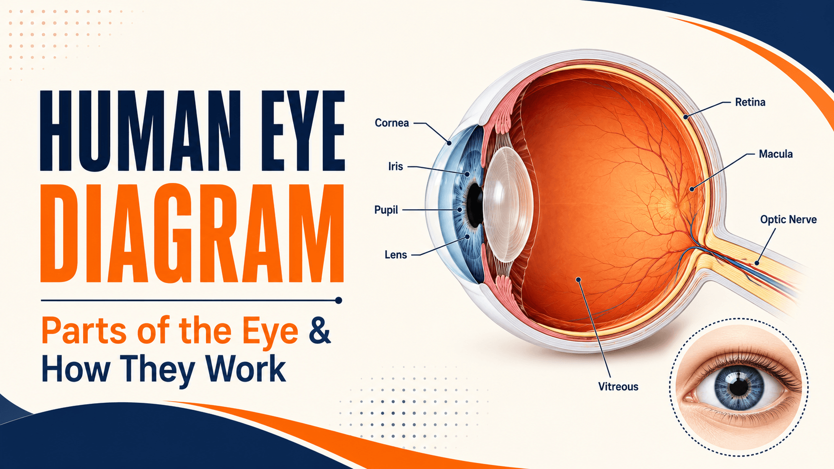
The main parts of the human eye are the cornea, sclera, conjunctiva, iris, pupil, lens, ciliary body, vitreous, retina, macula, choroid and optic nerve. Light passes through the cornea and pupil, is focused by the cornea and lens, and lands on the retina at the back of the eye. The retina converts light into electrical signals, and the optic nerve carries those signals to the brain, which interprets them as the image you see.
The Human Eye at a Glance
The eyeball sits in a bony socket called the orbit, cushioned by fat and moved by six small muscles. It is built in three layers:
- The outer layer is the tough white sclera, and at the front, the clear cornea
- The middle layer is the iris, ciliary body and choroid, which together control light, focusing and blood supply
- The inner layer is the retina, the light-sensitive tissue that lines the back of the eye
Inside, the eye is divided into fluid-filled spaces: the anterior chamber between the cornea and iris, the posterior chamber between the iris and lens, and the large vitreous cavity behind the lens, which makes up roughly two-thirds of the eyeball’s volume.
How Light Travels Through the Eye
Following the path of light is the simplest way to understand the diagram.
- Light enters through the cornea, the clear dome at the front, which does most of the focusing.
- It passes through the aqueous humour, the watery fluid in the front chamber.
- It passes through the pupil, the opening whose size the iris adjusts to control how much light gets in.
- The lens fine-tunes the focus, changing shape depending on whether you are looking near or far.
- It travels through the vitreous, the clear gel filling the back of the eye.
- It lands on the retina, where light-sensitive cells convert it into electrical signals.
- The optic nerve carries those signals to the brain, which assembles them into the image you experience.
The Front of the Eye
Cornea
The cornea is the clear, dome-shaped window at the front. It has no blood vessels, which is why it is transparent and gets its oxygen and nutrients from the tear film and the aqueous humour behind it.
Its main job is refraction, or bending light. The relaxed eye has a total focusing power of roughly 60 dioptres, and the cornea supplies about 40 of those, around two-thirds of the eye’s entire focusing power. Unlike the lens, its focus is fixed. Because its shape determines so much, small irregularities in curvature cause astigmatism, and progressive thinning causes keratoconus.
Sclera and conjunctiva
The sclera is the tough white outer coat that gives the eye its shape and protects its contents. The conjunctiva is a thin, transparent membrane covering the sclera and lining the inside of the eyelids. It is the tissue that becomes inflamed and red in conjunctivitis, and the layer beneath which a harmless red patch appears when a small surface vessel breaks.
Iris and pupil
The iris is the coloured ring, and the pupil is the black opening at its centre. The pupil is not a structure; it is simply a hole. The iris contains tiny muscles that widen the pupil in dim light and narrow it in bright light, with the opening varying from roughly 1.5 mm to 8 mm across.
If the two pupils are noticeably different sizes, that is called anisocoria, which is common and often harmless but occasionally signals a nerve problem.
Aqueous humour and the drainage angle
The ciliary body continuously produces a clear fluid called aqueous humour, which nourishes the cornea and lens and maintains the eye’s internal pressure. It drains away through a sieve-like structure called the trabecular meshwork, located in the angle where the iris meets the cornea.
This small structure matters more than its size suggests. When drainage cannot keep pace with production, pressure inside the eye rises the mechanism behind most glaucoma. Anatomy pages often skip this, but it is the single most important functional detail at the front of the eye.
The Focusing System
Lens
The crystalline lens sits behind the iris, roughly 10 mm across and 4 mm thick, and is transparent and flexible in youth. It supplies the remaining third of the eye’s focusing power and, unlike the cornea, it can change shape.
Two age-related changes affect it. From around the forties, it stiffens, making near focus harder presbyopia. Later, it can become cloudy, which is a cataract, treated by replacing it with an artificial lens.
Ciliary body and zonules
The ciliary body is a ring of muscle behind the iris. Fine fibres called zonules suspend the lens from it, like the strings holding a hammock. When the ciliary muscle contracts, tension on the zonules eases and the lens becomes rounder for near focus, a process called accommodation. The lens sits within a delicate membrane called the capsular bag, which is preserved during cataract surgery to hold the artificial lens in place.
Vitreous
The vitreous is the clear gel filling the space between the lens and retina. It is over 98% water and helps the eye keep its shape.
With age it liquefies and shrinks. Small clumps within it cast shadows seen as floaters, and when it separates from the retina, a posterior vitreous detachment, it can tug on the retina, causing flashes and occasionally a retinal tear or detachment.
The Back of the Eye
Retina
The retina is the light-sensitive tissue lining the back of the eye. Developmentally, it is an outgrowth of the brain.
It contains two kinds of photoreceptors. Rods are far more numerous, work in dim light, and handle peripheral vision but not colour. Cones work in bright light, provide colour vision and fine detail, and are concentrated in the centre. Damage to the retina from diabetic retinopathy is a leading cause of vision loss in adults.
Macula and fovea
The macula is the small central area of the retina responsible for sharp, detailed vision, reading, recognising faces, and seeing colour. At its centre is a tiny pit called the fovea, made up almost entirely of cones, which gives you your sharpest possible vision.
The macula is why you can read this line but cannot read the line above it without moving your eyes. Age-related macular degeneration affects exactly this area, which is why it damages central vision while side vision usually remains.
Optic nerve and the blind spot
The optic nerve is the cable carrying visual signals to the brain, made up of more than a million individual nerve fibres. Where it leaves the eye there are no photoreceptors at all, creating a genuine blind spot in each eye.
You never notice it, for two reasons: each eye covers the other’s blind spot, and the brain fills in the gap from surrounding information. Damage to this nerve causes permanent vision loss, since the fibres do not regenerate, the basis of optic atrophy and of glaucoma-related sight loss.
Choroid
The choroid is a layer rich in blood vessels sitting between the retina and sclera. It supplies oxygen and nutrients to the outer retina and absorbs stray light that would otherwise scatter and blur the image. Abnormal vessel growth from the choroid is what causes wet macular degeneration.
The Structures That Protect and Move the Eye
Not everything on an eye diagram is inside the eyeball.
- Eyelids and eyelashes shield the eye and spread tears with every blink.
- The tear film has three layers: an oily outer layer, a watery middle layer, and a mucous inner layer, and keeps the corneal surface smooth and clear. When it becomes unstable, the result is dry eye, and vision can blur between blinks.
- The lacrimal gland produces tears, which drain through small openings at the inner corner into the nose, which is why crying makes your nose run.
- Six extraocular muscles move each eye: four straight (rectus) muscles and two oblique muscles. They work as a coordinated pair across both eyes, and when that coordination fails, the result is a squint or double vision.
- The orbit, the bony socket, protects the eye from injury.
Parts of the Eye and Their Functions
|
Part |
What it does |
Commonly associated conditions |
|
Cornea |
Clear front window; provides about two-thirds of focusing power |
Astigmatism, keratoconus, corneal infection and scarring |
|
Sclera |
Tough white outer coat; maintains shape |
Scleritis, injury |
|
Conjunctiva |
Thin covering over sclera and inner eyelids |
Conjunctivitis, surface bleeding |
|
Iris |
Controls pupil size and light entering the eye |
Uveitis, anisocoria |
|
Pupil |
The opening through which light passes |
Reflects nerve and iris problems |
|
Aqueous humour |
Nourishes cornea and lens; maintains eye pressure |
Raised eye pressure |
|
Trabecular meshwork |
Drains aqueous fluid from the eye |
Glaucoma |
|
Lens |
Fine-tunes focus for near and distance |
Cataract, presbyopia |
|
Ciliary body |
Produces aqueous fluid; changes lens shape |
Affects focusing and eye pressure |
|
Vitreous |
Clear gel maintaining eye shape |
Floaters, vitreous detachment |
|
Retina |
Converts light into electrical signals |
Diabetic retinopathy, retinal detachment |
|
Macula and fovea |
Sharp central and colour vision |
Macular degeneration, macular hole |
|
Choroid |
Blood supply to the outer retina |
Wet macular degeneration |
|
Optic nerve |
Carries signals to the brain |
Glaucoma, optic atrophy, optic neuritis |
|
Tear film |
Keeps the corneal surface smooth and clear |
Dry eye |
|
Eye muscles |
Move and align the eyes |
Squint, double vision |
How the Eye Works Like a Camera
The comparison is genuinely useful, as long as you take it far enough.
- The cornea and lens together act as the camera lens, focusing incoming light.
- The iris and pupil work as the aperture, controlling how much light enters.
- The retina is the sensor, capturing the image.
- The optic nerve is the cable carrying data to the processor.
- The brain is the processor, and it does far more work than most people assume.
Here is the part the analogy usually leaves out: the image that lands on your retina is upside down and reversed. Your brain flips it. What you experience as seeing is not a picture arriving in your head but an interpretation your brain constructs, which is also why the blind spot goes unnoticed, and why vision problems can originate in the brain as well as the eye.
Where the analogy breaks down is repair. A camera lens can be swapped out. Most parts of the eye cannot; the retina and optic nerve in particular do not regenerate, which is why damage to them is usually permanent and why early detection matters so much.
Why Eye Anatomy Matters for Your Health
Understanding the diagram makes eye conditions far easier to follow, and it explains something patients often find puzzling: why a serious eye disease can produce no symptoms at all.
Conditions affecting the macula hit central vision first, so reading becomes difficult early. Conditions affecting the peripheral retina or optic nerve, such as glaucoma, take side vision first, and because the brain fills in gaps and each eye compensates for the other, people frequently notice nothing until a great deal of damage has occurred.
That is the practical reason a chart test alone is not enough. Checking the eye’s pressure, its drainage angle, its retina, and its optic nerve requires a proper examination, and a comprehensive eye check-up looks at each of these structures in turn.
See an eye specialist promptly if you notice sudden vision loss, a curtain or shadow across your vision, flashes with a sudden increase in floaters, eye pain with redness, or double vision; these point to specific structures needing urgent attention.
Conclusion
A human eye diagram becomes far more useful once each label carries a job. The cornea does the heavy focusing, the iris controls the light, the lens fine-tunes, the retina captures, and the optic nerve carries it all to a brain that does more interpretation than most people realise.
The practical value of knowing the layout is that it explains where things go wrong: cataract in the lens, glaucoma at the drainage angle and optic nerve, macular degeneration at the centre of the retina, dry eye on the surface. It also explains why several of these can advance silently.
If it has been more than a year or two since your last check, or you have noticed any change in your vision, book a comprehensive eye examination so each of these structures can be assessed properly.