



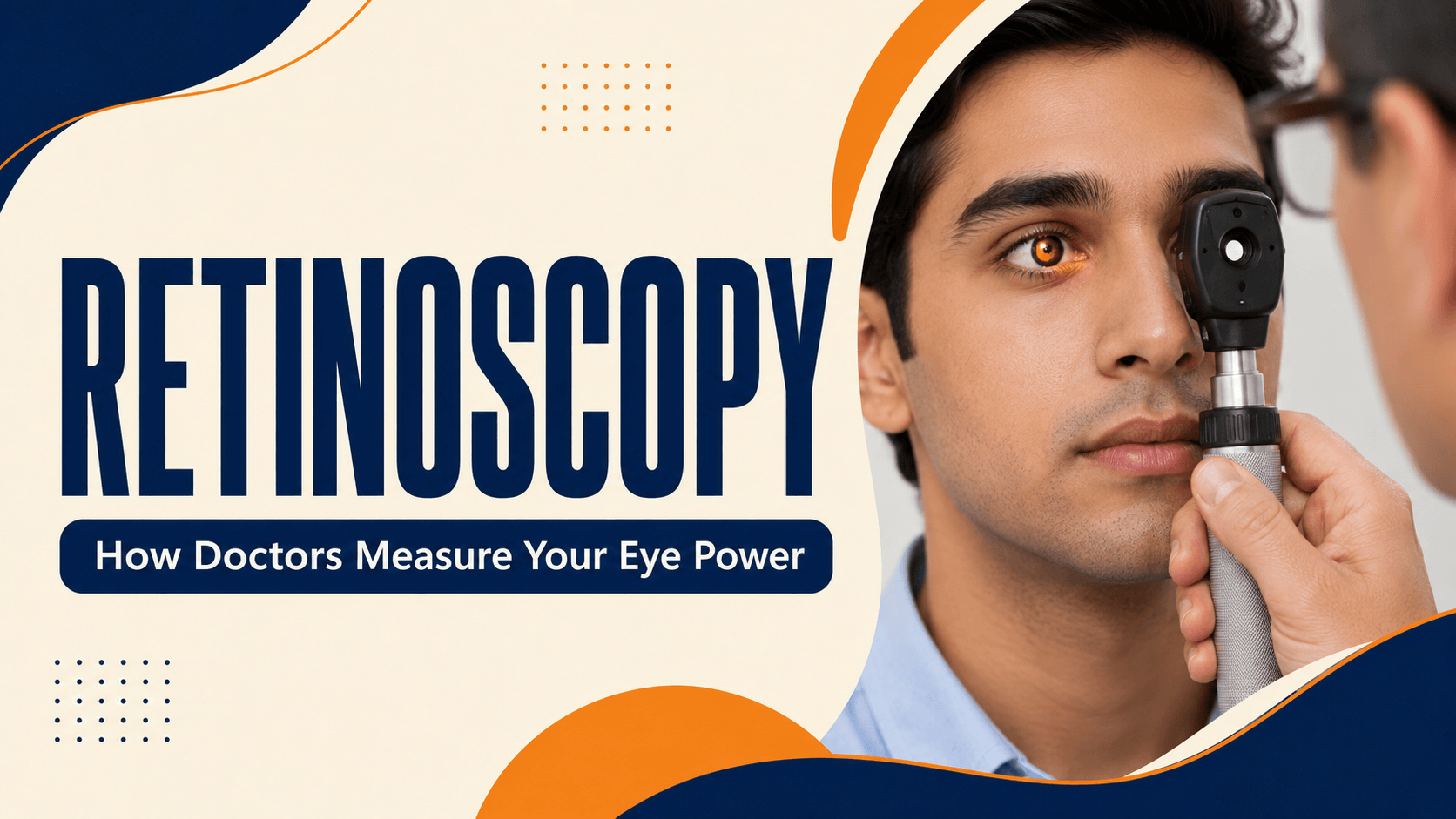
If you have ever had an eye examination where the doctor shone a light into your eye and flipped lenses in front of it without asking you a single question, you have had a retinoscopy.
It is one of the oldest techniques in eye care and still one of the most useful, particularly for children, who cannot reliably tell anyone how well they see.
Key Takeaway
Retinoscopy is a test that measures the eye’s focusing power by shining a light into the eye and observing how the light reflects back from the retina. The examiner places lenses in front of the eye until the reflection behaves in a particular way, which reveals the spectacle power needed. It is objective; it works without the patient answering anything, so it can be used on babies, young children and anyone unable to respond.
What Is Retinoscopy?
Retinoscopy, sometimes called skiascopy, is a method of measuring refractive error, the technical name for what most people call eye power or spectacle number. It shows whether an eye is short-sighted, long-sighted or has astigmatism, and by how much.
The examiner uses a handheld instrument called a retinoscope, which projects a narrow beam of light into the eye. Most modern instruments project a stripe of light, which is why the technique is often called streak retinoscopy.
The defining feature is that it is an objective test. You are not asked to compare lenses or read a chart. The answer comes from what the examiner observes, not from what you report.
What the Examiner Is Actually Looking At
When light enters your eye, some of it bounces back off the retina at the back. Through the retinoscope, the examiner sees this returning light as a glow in your pupil, the same effect that makes eyes look red in flash photographs.
As the examiner sweeps the light beam across your eye, that glow appears to move. The direction and speed of that movement depend on your eye’s focusing power.
The examiner then holds different lenses in front of the eye. Each lens changes how the glow moves. When the correct lens is reached, the movement stops and the pupil fills with light evenly. That lens value, adjusted for the examiner’s distance from you, gives your eye power.
It looks effortless when done well. It is a skilled clinical technique that takes considerable training.
Why Retinoscopy Still Matters When Machines Can Measure Eye Power
Most opticals and clinics in India have an autorefractor the machine you rest your chin on, look at a picture inside, and get a printed reading from. It is quick and useful. It is not the whole examination.
A printed autorefractor reading is a starting point, not a prescription.
|
Retinoscopy |
Autorefraction |
Subjective refraction |
|
|
How it works |
Examiner observes light reflected from the retina and neutralises it with lenses |
Machine measures the eye automatically |
Patient compares lens options and reports which is clearer |
|
Needs your responses? |
No |
No |
Yes |
|
Works on babies and young children |
Yes |
Only if the child can stay still and look steadily |
No |
|
Works with cataract or a hazy cornea |
Often yes, with skill |
Frequently unreliable |
Limited |
|
Main limitation |
Depends on examiner skill |
Can over-read focusing effort, especially in children |
Needs an alert, cooperative patient |
In practice, a proper refraction usually combines them: an objective starting measurement from retinoscopy or the machine, then subjective refinement where the patient is old enough to take part.
Where the difference matters most is in children. A young eye can focus hard without the child being aware of it. An autorefractor reading taken while that focusing effort is active can suggest more short-sightedness than really exists, which can mean a child is given a stronger spectacle power than they need. This is the main reason a machine reading alone is not enough for a child, and why proper paediatric testing involves drops and a trained examiner.
Who Needs Retinoscopy?
Retinoscopy is particularly valuable for:
- Babies and young children, who cannot answer questions about their vision
- Children being checked for squint or lazy eye, where an accurate power measurement is essential to treatment
- People with intellectual disability, dementia or communication difficulty, or anyone unable to respond reliably
- Patients with very high spectacle powers, where small errors matter
- Eyes with an irregular cornea, such as in keratoconus or after injury
- Patients where the machine reading and the symptoms do not match
- Adults before cataract surgery or refractive surgery, as part of a full assessment where accuracy is critical
It is also simply part of a thorough eye examination for many adults, even when everything is straightforward.
What Happens During a Retinoscopy Test
The test itself is quick, usually a few minutes per eye.
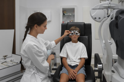
- You sit facing the examiner in a dimly lit room. Low lighting makes your pupil larger naturally and the reflection easier to see.
- You are asked to look at a distant target — often a letter chart or a light across the room and to keep looking past the examiner rather than at their light.
- The examiner shines the retinoscope beam into one eye and moves it slowly across, watching the glow in your pupil.
- Lenses are held in front of the eye, either in a trial frame or on a handheld bar, and changed one by one.
- The examiner watches how each lens changes the movement of the light until it neutralises.
- The other eye is measured the same way.
- If you are old enough to take part, the result is then fine-tuned by asking you to compare lens choices.
For a small child, a parent usually holds them on their lap. Toys, a video or a moving light are often used to hold the child’s attention. Children rarely find it distressing — from their side, someone is simply showing them a light.
Why Children Usually Need Eye Drops First
This is the part that most worries parents, and it has a clear explanation.
Your eye contains a small muscle that changes the lens shape to focus on near objects. In children this muscle is strong and constantly active, and a child cannot switch it off on request. If it is working during the test, the measurement reflects the focusing effort rather than the eye’s true resting power.
To get an accurate result, the examiner uses cycloplegic eye drops, which temporarily relax that focusing muscle and also widen the pupil. Retinoscopy done after these drops is often called wet retinoscopy, and the process as a whole is called cycloplegic refraction.
This is not an optional extra for children. It is what makes the number reliable, and an unreliable number means the wrong glasses.
The drops sting briefly. That is the honest answer to give a child, and warning them is better than surprising them. Then there is usually a wait of around 30 to 45 minutes while the drops take effect, and the examiner may check that the pupils have stopped reacting to light before starting.
One point relevant to Indian patients: darker brown irides can need more time, or an extra drop, for full effect than lighter eyes. If your child’s appointment takes longer than you expected, this is often why.
How Long the Drops Last and What to Plan For
Nobody tells patients this, and it is the most practically useful part of the whole appointment.
After cycloplegic drops, expect:
- Enlarged pupils for several hours
- Blurred near vision — reading, writing and phone screens will be difficult
- Sensitivity to bright light, which can be uncomfortable outdoors
How long depends on which drop is used. Shorter-acting drops typically wear off within several hours; the commonly used cycloplegic agents can affect focusing for the rest of the day and sometimes into the next. Longer-acting drops, used in specific situations, last considerably longer. Ask which drop was used and how long its effect is expected to last — the answer changes what you should plan.
Practical steps that make the day easier:
- Do not plan for your child to return to school that afternoon, or for exams, homework or detailed close work.
- Bring sunglasses or a cap for the journey home. Bright Indian daylight after dilation is genuinely uncomfortable.
- Do not drive yourself home if you are the one having the drops. Arrange a companion or transport.
- Allow far more time than the test needs. With waiting for the drops to work, budget a couple of hours at the hospital rather than a few minutes.
- Bring something to occupy a child during the waiting period.
Vision returns to normal on its own. No treatment is needed to reverse the drops.
Is Retinoscopy Safe? Does It Hurt?
Retinoscopy itself is completely painless and carries no risk. Nothing touches your eye. The light is bright but harmless, and the test is over in minutes.
The dilating drops have mild, temporary effects: stinging on instillation, blurred near vision and light sensitivity, as described above. These are expected effects rather than complications.
Two genuine cautions apply to the drops rather than the test:
- Some adults have a narrow drainage angle inside the eye, where dilation carries a small risk of raising eye pressure. Your ophthalmologist checks for this before dilating.
- Cycloplegic drops occasionally cause flushing, dryness of the mouth, restlessness or drowsiness, particularly in small children and at higher strengths. Tell the clinic if your child has reacted to eye drops before, and report any unusual symptoms after leaving.
Mention any allergy, any previous reaction to eye drops, and any neurological or heart condition before the drops are given.
What Happens After the Test
The retinoscopy result is one part of an examination, not the whole conclusion.
- If glasses are needed, you will be given a prescription. For children, doctors often prescribe slightly differently from the raw cycloplegic figure, allowing for the fact that the focusing muscle was relaxed during measurement. A number that looks different from a shop’s machine reading is not necessarily an error.
- If a squint or lazy eye is found, correcting the refractive error is usually the first step, and further treatment such as patching may follow under paediatric eye care.
- If the eye examination raises other findings, further tests may be arranged.
- A follow-up is usually scheduled, since children’s eye power changes as they grow.
Children with glasses generally need review at least once a year, and more often if the power is changing quickly or amblyopia is being treated.
When to Get Your Child’s Eyes Tested
Children rarely complain about poor vision, because they have no basis for comparison. Parents usually notice behaviour before the child reports a problem.
Arrange an eye test if your child:
- Sits very close to the television, or holds books and screens close to the face
- Squeezes or narrows the eyes to see clearly
- Tilts or turns the head to look at things
- Rubs the eyes often, or complains of headaches after school
- Has one eye that drifts inward or outward, even occasionally
- Is doing worse at school, or avoiding reading
- Has a family history of spectacles from a young age, squint or lazy eye
Seek assessment promptly for any visible misalignment of the eyes, a white or unusual reflection in the pupil in photographs, or a sudden change in vision. These need examination rather than waiting for a routine check.
Because amblyopia responds best when treated early in childhood, a delayed diagnosis matters. A comprehensive eye test is the way to establish what is actually going on.
Conclusion
Retinoscopy is a quick, painless and remarkably capable test. By reading light reflected from the back of the eye, a trained examiner can determine spectacle power in someone who cannot say a word about their vision — which is exactly why it remains central to children’s eye care.
If your child has been advised retinoscopy with dilating drops, the two things worth knowing are that the drops are what make the result trustworthy, and that the rest of the day should be planned around blurred near vision and light sensitivity. If you have noticed any of the signs described above, book an appointment rather than waiting for a school screening.