



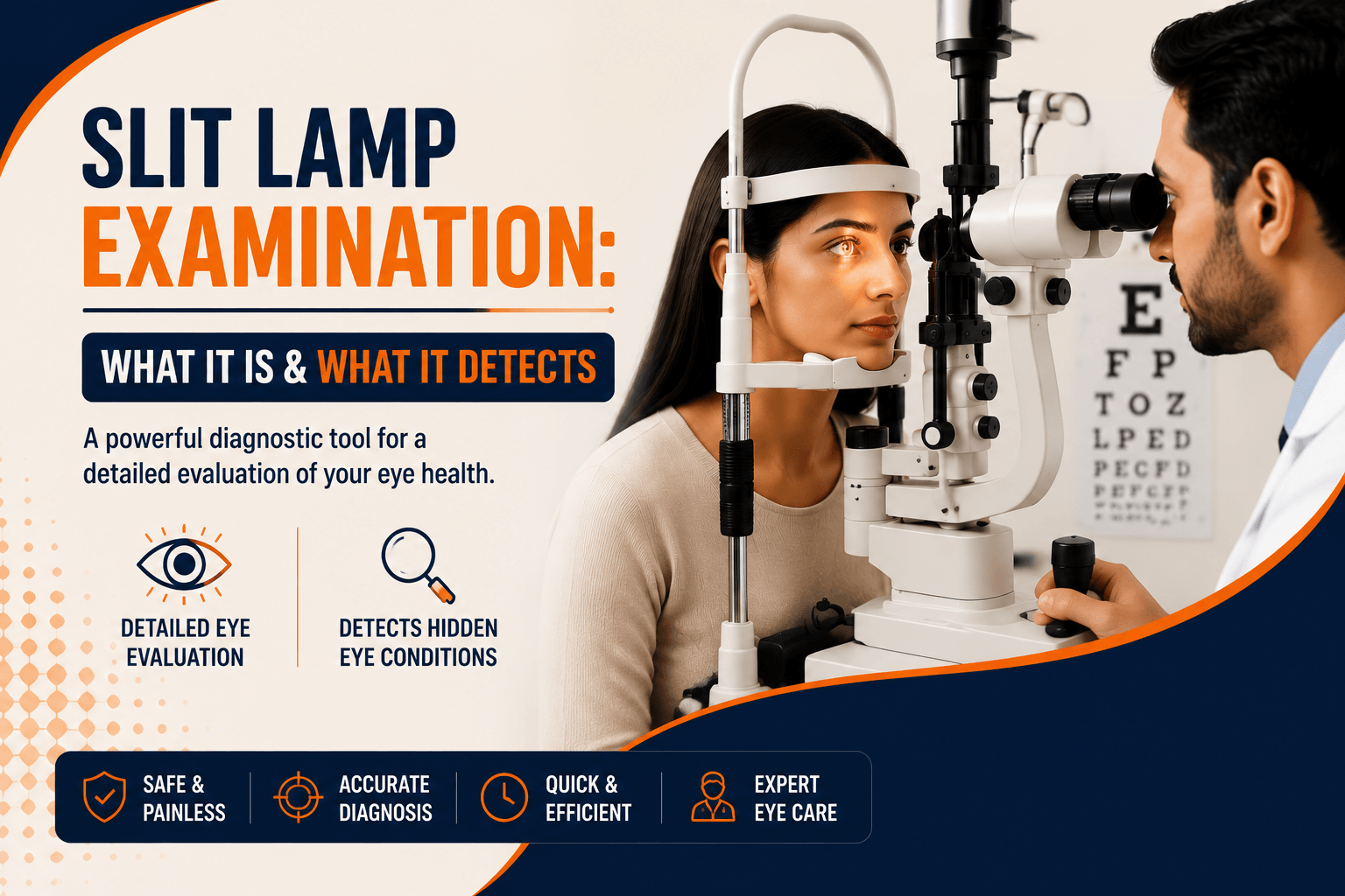
If you have ever rested your chin on a padded support while a doctor shone a narrow, very bright beam into your eye, you have had a slit lamp examination.
It is the single most-used instrument in eye care, and most patients go through it repeatedly without anyone explaining what it is doing.
Key Takeaway
A slit lamp examination uses a special microscope and a narrow beam of light to examine the structures of your eye under high magnification. It shows the eyelids, the surface of the eye, the cornea, the fluid-filled chamber behind it, the iris and the lens, and with an added lens, the retina at the back. The examination itself is painless and takes only a few minutes.
What Is a Slit Lamp Examination?
A slit lamp is a microscope combined with an adjustable light source, mounted on a table with a chin rest and forehead band. The medical term for the examination is biomicroscopy.
The instrument magnifies your eye, typically somewhere between about six and forty times, and the examiner adjusts that magnification depending on what they are looking at.
What makes it powerful is not just magnification. It is the shape of the light.
Why the Light Is a Slit
Shine a wide light at the eye and you see a surface. Narrow that light into a thin slit and angle it, and something more useful happens: the beam cuts an optical section through the transparent tissues, rather like slicing a thin section through a piece of glass.
That is what lets an examiner see into the cornea rather than just across it, assessing its separate layers, and judging the depth of a scratch, an ulcer or an opacity rather than only its position.
The examiner can widen or narrow the beam, change its height, angle it from either side, and switch between filters. Each setting reveals something different.
What a Slit Lamp Examination Detects
This is where the instrument earns its place. Working from the front of the eye backwards:
|
Part of the eye |
What the examiner is looking for |
|
Eyelids and lashes |
Inflammation of the lid margins as in blepharitis, blocked oil glands, a stye, lid position problems, lash abnormalities |
|
Conjunctiva (the clear membrane over the white) |
Redness patterns, discharge, the follicles and papillae that help distinguish infective from allergic conjunctivitis, growths |
|
Tear film |
Signs of dry eye, including how quickly the tear film breaks up and where the surface is damaged |
|
Cornea (the clear front window) |
Scratches and abrasions, foreign bodies, infection and corneal ulcers, scarring, swelling, thinning or irregular shape as in keratoconus |
|
Anterior chamber (fluid space behind the cornea) |
Inflammatory cells indicating uveitis, blood, and how deep or shallow the space is relevant to certain kinds of glaucoma |
|
Iris (the coloured part) |
Inflammation, adhesions, irregularities, growths |
|
Lens |
Cloudiness and its pattern; this is how a cataract is identified and graded |
|
Retina and optic nerve (with an added lens) |
Changes from diabetic retinopathy, swelling such as macular oedema, optic nerve appearance, retinal tears |
Contact lens wearers get particular value from it. The slit lamp shows how a lens sits and moves on the eye, and picks up the early surface changes that lens wear can cause often before the wearer notices anything.
What Happens During the Test
The examination is quick and requires nothing from you except sitting still.
- You sit facing the instrument in a dimmed room and rest your chin on the support, with your forehead against the band.
- The examiner adjusts the height so your eyes align with the marker on the frame.
- You are asked to look straight ahead, or in particular directions, and to blink normally between looks.
- A narrow beam is directed at your eye and moved across it while the examiner looks through the microscope.
- The beam and magnification are adjusted repeatedly as different structures are examined.
- Your eyelid may be gently held or everted to see the surface underneath.
- The other eye is examined the same way.
The whole thing usually takes a few minutes per eye. The light is bright and can feel dazzling, but it causes no harm and you may see an afterimage for a short while.
The Orange Drop and the Blue Light
This is the part patients most often ask about afterwards.
Fluorescein is a harmless orange-yellow dye, applied either as a drop or from a small paper strip touched briefly to the eye. Under the slit lamp’s cobalt blue filter, it glows bright green-yellow.
The reason it is useful is elegant. Fluorescein does not stick to a healthy, intact eye surface. Where the surface layer is broken a scratch, an ulcer, an area of dryness the dye settles into the gap and lights up under blue light, showing the examiner the exact size and shape of the damage.
It is also used during eye pressure measurement, for a different reason explained below.
The dye washes out naturally in your tears within a short time. It may tint your tears briefly and can stain soft contact lenses, which is one reason you are asked to remove lenses first.
When Something Does Touch Your Eye
You may read elsewhere that nothing touches your eye during this test. That is true of the slit lamp examination itself but not always of the tests done at the same machine, and it is worth knowing in advance.
- Eye pressure measurement (applanation tonometry). The most accurate method uses a small prism mounted on the slit lamp that rests very gently against the front of the cornea for a second or two. Anaesthetic drops are put in first, usually combined with fluorescein, so you feel pressure at most, not pain. Many people do not realise contact has happened.
- Examining the drainage angle (gonioscopy). A special contact lens is placed on the anaesthetised eye to view the angle where fluid drains important in assessing glaucoma risk.
- Fluorescein strips, where the dye is applied with a paper strip rather than a dropper.
None of these is painful, and all are done after numbing drops where contact is involved. But being told “nothing will touch your eye” and then experiencing contact is unsettling, so it is better to know.
Does the Slit Lamp Replace Other Eye Tests?
No, and this is a common misunderstanding. The slit lamp is the platform on which much of an eye examination happens, but it does not do everything.
|
What it does |
What it does not do |
|
Examines eye structures in magnified detail |
Measure your spectacle power that needs refraction |
|
Serves as the base for eye pressure measurement |
Maps your field of vision that needs a visual field test |
|
Allows a detailed retinal view with an added lens |
Produces cross-sectional scans of the retina or optic nerve that need an OCT scan |
|
Grades cataract and assesses the eye surface |
Photographs the retina for records or screening that needs fundus imaging |
A full assessment usually combines several of these. If your appointment involved more than one machine, that is why each answers a different question, and the slit lamp answers the ones about structure and appearance.
Will You Need Dilating Drops?
Not always. The front of the eye can be examined perfectly well without them.
Dilating drops are used when the examiner needs a proper view of the back of the eye the retina and optic nerve through an added lens. This is standard for diabetes screening, for anyone with retinal symptoms, and for many routine comprehensive checks.
If drops are used, expect:
- Enlarged pupils and blurred near vision for several hours
- Sensitivity to bright light
- No ability to read comfortably or drive safely until it wears off
Bring sunglasses, arrange transport rather than driving yourself, and allow extra time at the hospital. Ask which drop was used, as the duration varies.
Is It Safe? Does It Hurt?
The slit lamp examination is painless and very safe. It uses ordinary visible light, no radiation, no lasers during a standard examination, and no injections.
What people do experience:
- Dazzle from the bright light, and an afterimage lasting a minute or two
- A reflex urge to blink, which is normal and expected
- Mild dryness or a gritty feeling afterwards, from keeping the eyes open under a warm light
- Brief stinging from anaesthetic or fluorescein drops, if used
- Blurred vision and light sensitivity for hours, if dilating drops were used
People with a painful eye or marked light sensitivity may find the examination harder to tolerate. So the examiner can work faster, use lower light, or use anaesthetic drops to make it manageable.
Very occasionally someone reacts to the drops with redness or itching. Mention any previous reaction to eye drops before the test.
Why You Have This Test at Almost Every Visit
Because the eye changes, and because the slit lamp is how an eye doctor actually looks at it.
- It is the baseline. Comparing today’s appearance with last visit’s is how progression is judged: whether a cataract is denser, an ulcer is healing, or inflammation has settled.
- Treatment is monitored through it. Whether drops are working shows up on the eye surface before it shows up in how you feel.
- Many conditions are silent early on. Surface damage, early cataract, and quiet inflammation frequently cause no symptoms at all.
- It guides what else is needed. What the examiner sees determines whether you need imaging, pressure checks or further tests.
Repeating the examination is not duplication. It is the comparison that carries the information.
When to Get Your Eyes Examined
Book an appointment if you have:
- Redness that is not settling, or discharge
- A gritty or foreign-body sensation that persists
- Blurred vision, or vision that fluctuates
- Discomfort while wearing contact lenses
- Diabetes and no eye check in the past year
- A family history of glaucoma
Seek same-day care for eye pain, sudden reduced vision, marked light sensitivity, a red painful eye while wearing contact lenses, a visible white or grey spot on the eye, or any eye injury, particularly one involving a chemical, plant material or a high-speed particle.
A comprehensive eye test includes the slit lamp examination alongside whichever other tests your symptoms call for.
Conclusion
The slit lamp is the instrument an eye doctor reaches for first, and for good reason: a thin beam of light and a good microscope reveal more about the front of the eye than any scan. It is quick, painless, and does much of the work of diagnosing eye disease.
The parts worth knowing are the small ones. The orange dye finds breaks in the eye’s surface. The blue light makes them glow. And if something briefly touches your eye during pressure measurement, that is expected, and numbing drops mean you will barely feel it.
If you have redness, discomfort, blurring or any symptom that has not settled, that is exactly what this examination is for. Book an appointment and have your eyes properly looked at.